. 2024 Jan 19;22(1):57.
doi: 10.1186/s12964-024-01471-7.

Extracellular lipidosomes containing lipid droplets and mitochondria are released during melanoma cell division

Affiliations

Extracellular lipidosomes containing lipid droplets and mitochondria are released during melanoma cell division

Jana Karbanová et al. Cell Commun Signal. .

Abstract

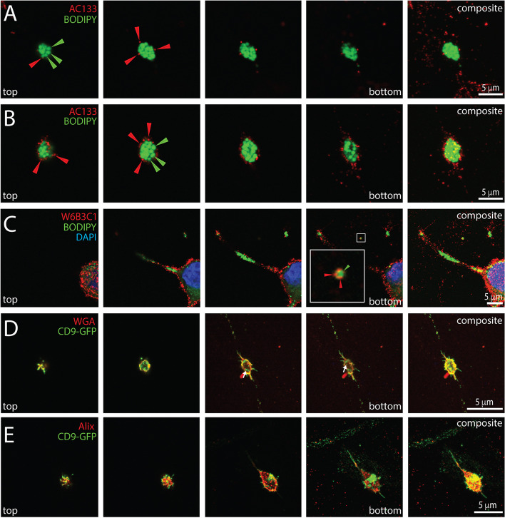
Background: The incidence of melanoma is increasing worldwide. Since metastatic melanoma is highly aggressive, it is important to decipher all the biological aspects of melanoma cells. In this context, we have previously shown that metastatic FEMX-I melanoma cells release small (< 150 nm) extracellular vesicles (EVs) known as exosomes and ectosomes containing the stem (and cancer stem) cell antigenic marker CD133. EVs play an important role in intercellular communication, which could have a micro-environmental impact on surrounding tissues.

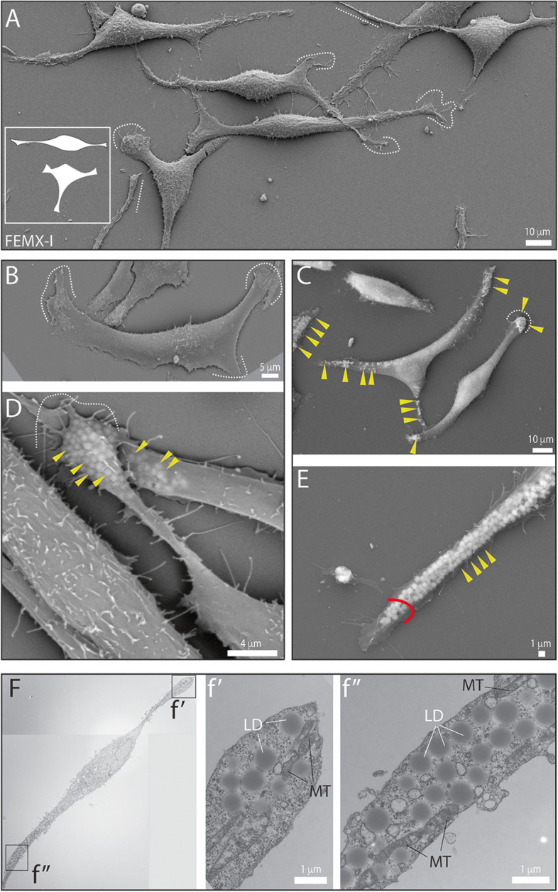
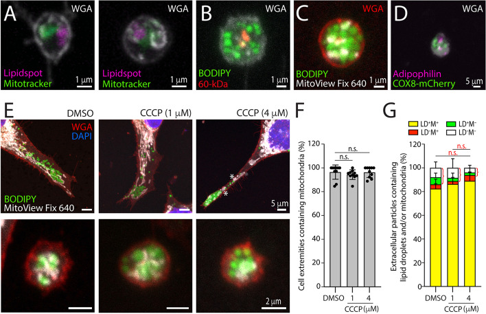
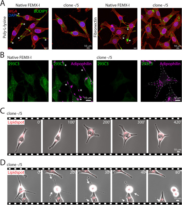
Results: We report here a new type of large CD133+ EVs released by FEMX-I cells. Their sizes range from 2 to 6 µm and they contain lipid droplets and mitochondria. Real-time video microscopy revealed that these EVs originate from the lipid droplet-enriched cell extremities that did not completely retract during the cell division process. Once released, they can be taken up by other cells. Silencing CD133 significantly affected the cellular distribution of lipid droplets, with a re-localization around the nuclear compartment. As a result, the formation of large EVs containing lipid droplets was severely compromised.

Conclusion: Given the biochemical effect of lipid droplets and mitochondria and/or their complexes on cell metabolism, the release and uptake of these new large CD133+ EVs from dividing aggressive melanoma cells can influence both donor and recipient cells, and therefore impact melanoma growth and dissemination.

Keywords: Cell division; Extracellular vesicle; Lipid droplet; Melanoma; Mitochondrion; Prominin-1.


Conflict of interest statement

The authors declare no competing interests.

Figures

Fig. 1
Fig. 1
Melanoma FEMX-I cells harbor bipolar or triangular morphotypes and contain lipid droplets and mitochondria at their extremities. A-F FEMX-I cells growing on poly-L-lysine-coated coverslips (A-E) or on plastic Petri dishes (F) were processed for scanning (A-E) and transmission (F) electron microscopy. Two main morphotypes are observed; cells with a bipolar and tripolar shape (see illustration in A). Their extremities are either short and flattened with filopodia adhering to the support (A-D, dashed line) or long and narrow (A, E, straight dotted line and red arc). Various small, rounded particles are observable particularly at the extremities of the cells when applying a higher acceleration voltage (15 kV instead 5 kV) during SEM analysis (C-E, yellow arrowhead). These homogeneous intracellular particles appear as opaque spheres by TEM, which is the typical appearance of lipid droplets (LD, F, see enlargement of images f’ and f’’). Mitochondria (MT) are present in the vicinity of lipid droplets (see also Additional file 1: Fig. S2). Scale bars are indicated
Fig. 2
Fig. 2
Fluorescence detection of lipid droplets and mitochondria at the extremities of melanoma cells. A-F Native FEMX-I cells (A-E) or COX-8-mCherry-transfected cells (F) growing either on poly-L-lysine (A) or fibronectin-coated (B-F) supports were processed for CLSM (A, C-F) or live-cell phase-contrast/fluorescence video microscopy (B). PFA-fixed and saponin-permeabilized cells were immunolabeled with an anti-adipophilin (A, F) or anti-60-kDa mitochondrial antigen (E) antibody and/or stained with fluorescent dyes BODIPY™ 493/503 (A, D, E) or LipidSpot™ 610 (B, C). Alternatively, cells were co-stained with MitoTracker™ Red CMXRos (C) or MitoView™ Fix 640 (D). Cells were counterstained with DAPI (A, C, blue) or fluorescence-conjugated WGA (C, F) to highlight nuclei and glycoconjugates at the cell membrane, respectively. LipidSpot-stained cells were observed alive and elapsed time in minutes is shown on the top-right corner (B). Samples were pseudo-colored with a given marker as indicated. The images presented in panel B are excerpted from the Additional file 2: Video S1. Asterisks indicate the enrichment of lipid droplets at the cell extremities (A, B), while the symbol @ shows a cell changing from a bipolar to a tripolar morphology over time (B). The arrow points to a small EV containing lipid droplets (A). DIC, differential interference contrast. Scale bars are indicated
Fig. 3
Fig. 3
Cell extremities during cell division and extracellular lipidosomes. A-J FEMX-I (A-F, J) or CD9-deficient FEMX-I (shCD9, G-I) cells growing on poly-L-lysine-coated glass coverslips were processed for SEM using 5-kV (A, B and F, left panel) or 15-kV (C-E, F, right panel, G-J) accelerating voltage. Note the presence of large lipid droplet-filled membrane structures (e.g., numbered 1–3 in B) connected with a membrane bridge containing numerous filopodia (A-C, E, dashed lines) to rounded cell body during cell division (A-H, blue line). Filopodia are barely present in shCD9 cells (G-I). Cell extremities can detach completely from the cells, resulting in the formation of larger EVs containing various lipid droplets (I, J). Some regions of interest indicated by colored boxes (A, B, D, G) have been enlarged (A, E, F, H) as indicated. The yellow arrowhead points to a lipid droplet, while the white arrow points to a microvillar-like structure on extracellular lipidosomes. The symbol # indicates a membrane rupture during sample preparation. Scale bars are indicated
Fig. 4
Fig. 4
Biogenesis of extracellular lipidosomes occurs during cell division or cell migration. A-D FEMX-I cells growing on fibronectin-coated supports were recorded in live by phase-contrast/fluorescence video microscopy after staining with LipidSpot™ 488/610. During cell division, cells can reabsorb material from their extremities (A, dashed line and white arrow) or lose it, resulting in the formation of extracellular lipidosomes (B, white arrowhead). During migration, cells can release extracellular lipidosomes (C, D, white arrowhead). Extracellular lipidosomes can be either large (B, C) or small (D). The very thin process linking an extracellular lipidosome to the donor cell can withstand traction before breaking (C, yellow bracket). Red arrow indicates the orientation of cell migration. Elapsed time in minutes is shown on the top-right corner. The images are excerpted from the Additional file 4: Video S3 (A, top), Additional file 6: Video S5 (B), Additional file 7: Video S6 (C) and Additional file 8: Video S7 (D). Scale bars are indicated
Fig. 5
Fig. 5
Extracellular lipidosomes are taken up by non-migrating and migrating cells. A, B FEMX-I cells growing on fibronectin-coated supports were recorded in live by phase-contrast/fluorescence video microscopy after staining with LipidSpot™ 610. Non-migrating (A) and migrating (B, arrow) cells can uptake an extracellular lipidosome (asterisk). The latter can be detected inside the recipient cells (A, arrowhead). Elapsed time in minutes is shown on the top-right corner. The images are excerpted from the Additional file 9: Video S8 (A) and Additional file 10: Video S9 (B). Scale bars are indicated
Fig. 6
Fig. 6
Cytoskeleton components associated with extracellular lipidosomes. A-C FEMX-I cells growing on fibronectin-coated supports were processed for CLSM. PFA-fixed and saponin-permeabilized cells were immunolabeled with anti-Vimentin, Nestin (A) or α-tubulin (C) antibodies or stained with fluorochrome-conjugated phalloidin (B). All samples were co-stained with the fluorescent dye BODIPY™ 493/503 and counterstained with DAPI to highlight lipid droplets and nuclei, respectively. Composite images of all x–y optical sections are shown. Cell extremities and extracellular lipidosomes are displayed in the left and right panels, respectively. The yellow bracket indicates the very thin Nestinprocess linking an extracellular lipidosome to the donor cell prior rupture (A). PC, phase contrast image. Scale bars are indicated
Fig. 7
Fig. 7
Expression of integrins at cell extremities and extracellular lipidosomes. A-F FEMX-I cells growing on fibronectin-coated supports were processed for CLSM. PFA-fixed and saponin-permeabilized cells were immunolabeled with various anti-integrin (ITG) antibodies as indicated and co-stained with fluorescent dyes LipidSpot™ 610 and fluorescence-conjugated WGA to highlight lipid droplets and glycoconjugates, respectively. Samples were counterstained with DAPI to highlight nuclei. Composite images of all x–y optical sections are shown and a given marker was pseudo-colored as indicated. Cells and extracellular lipidosomes are shown in the left and right panels, respectively, except for those in panel A’-F’. The yellow bracket indicates the very thin ITG β1+ process linking an extracellular lipidosome to the donor cell prior rupture, while arrowhead indicates specific ITG at cell extremities. Scale bars are indicated
Fig. 8
Fig. 8
Extracellular lipidosomes contain mitochondria. A-D Native (A-C) or COX-8-mCherry-transfected (D) FEMX-I cells growing on fibronectin-coated supports were processed for CLSM. Cells preincubated with MitoTracker™ Red CMXRos (A) or MitoView™ Fix 640 (C) were PFA-fixed and co-stained with fluorescent dyes LipidSpot™ 610 (A) or BODIPY™ 493/503 (C) or saponin-permeabilized, immunolabeled with an anti-60-kDa mitochondrial antigen antibody and stained with BODIPY™ 493/503 (B). Alternatively, COX-8-mCherry-transfected cells were permeabilized and immunolabeled with an anti-adipophilin antibody (D). Samples were also co-stained with fluorescence-conjugated WGA to highlight glycoconjugates at the cell membrane. EG Native FEMX-I cells were incubated without (DMSO, control) or with 1 or 4 µM of CCCP for 8 h and for the 2 last hours were additionally co-incubated with MitoView™ Fix 640. After fixation and staining with BODIPY™ 493/503, cells were co-stained with fluorescence-conjugated WGA and DAPI to highlight and glycoconjugates at the cell membrane and nuclei, respectively. In all cases, composite images of all x–y optical sections are shown and a given marker was pseudo-colored as indicated. Cell extremities (upper panels) and the extracellular lipidosomes (lower panels) are shown (E). Note that in drug-treated cells, mitochondria are rounded (asterisk). Mitochondria-containing and mitochondria-free cell extremities (F) and extracellular particles (G) were quantified under various conditions as indicated. More than 200 cell extremities and 100 extracellular particles per condition were analyzed from 2 and 3 independent experiments, respectively. Each individual data in panel F corresponds to the percentage of cell extremities found in an area of 0.056 mm2. Extracellular particles were classified as containing or not lipid droplets (LD) and mitochondria (M), as indicated (G). Mean ± S.D. are presented. Although not significant, a slight reduction in extracellular particles without mitochondria (green and white, red bracket) is observed after incubation with CCCP. N.s. not significant, Scale bars are indicated
Fig. 9
Fig. 9
Detection of CD133 on melanoma cell-derived extracellular lipidosomes. A-E Native (A-C) or CD9-GFP-transfected (D, E) FEMX-I cells growing on poly-L-lysine coverslips were processed for CLSM. PFA-fixed cells without (A-D) or with (E) saponin-permeabilization were immunolabeled with an anti-CD133 antibody (clone AC133 or W6B3C1) and co-stained with BODIPY™ 493/503 (A-C). Alternatively, they were either stained with fluorescence-conjugated WGA (D), which labels glycoconjugates at the cell membrane, or immunolabeled for Alix (E) and co-observed with CD9-GFP. Cells were counterstained with DAPI to highlight nuclei. Single x–y optical sections from top to bottom (four panels on the left) and composite (right panel) images of all sections are shown. A small CD133+ EV containing few lipid droplets is enlarged in panel C. Red and green arrowheads indicate CD133 and lipid droplets, respectively, while white arrow points CD9-GFP inside the large EVs. Scale bars are indicated
Fig. 10
Fig. 10
Silencing CD133 affects the distribution of lipid droplets and impedes the biogenesis of extracellular lipidosomes. A-D Native or CD133-deficient (clone –/5) FEMX-I cells growing either on poly-L-lysine (A) or fibronectin-coated (A-D) supports were either processed for CLSM (A, B) or recorded in live by phase-contrast/fluorescence video microscopy after staining with LipidSpot™ 610 (C, D). For CLSM, PFA-fixed cells without (A) or with (B) saponin-permeabilization were co-stained with BODIPY™ 493/503 and fluorescence-conjugated WGA (A) to highlight lipid droplets and glycoconjugates at the cell membrane, respectively, or double-immunolabeled with antibodies directed against CD133 (293C3) and adipophilin (B). For the live cell imaging, elapsed time in minutes is shown on the top-right corner. The images are excerpted from the Additional file 11: Video S10 (C) and Additional file 12: Video S11 (D). Note the EVs lacking lipid droplets during cell division of CD133-deficient cells (D, arrowhead). Asterisks indicate the concentration of lipid droplets at cell extremities of native cells (A, B), while arrows indicate the retraction of cell extremities (D). The dashed lines mark the outline of the CD133-deficient cells (B). Scale bars are indicated

References

    1. Arnold M, Singh D, Laversanne M, Vignat J, Vaccarella S, Meheus F, et al. Global burden of cutaneous melanoma in 2020 and projections to 2040. JAMA Dermatol. 2022;158(5):495–503. doi: 10.1001/jamadermatol.2022.0160. - DOI - PMC - PubMed
    1. Bedrosian I, Faries MB, Guerry D, 4th, Elenitsas R, Schuchter L, Mick R, et al. Incidence of sentinel node metastasis in patients with thin primary melanoma (< or = 1 mm) with vertical growth phase. Ann Surg Oncol. 2000;7(4):262–267. doi: 10.1007/s10434-000-0262-z. - DOI - PubMed
    1. Rinderknecht M, Detmar M. Tumor lymphangiogenesis and melanoma metastasis. J Cell Physiol. 2008;216(2):347–354. doi: 10.1002/jcp.21494. - DOI - PubMed
    1. Damsky WE, Rosenbaum LE, Bosenberg M. Decoding melanoma metastasis. Cancers (Basel) 2010;3(1):126–163. doi: 10.3390/cancers3010126. - DOI - PMC - PubMed
    1. Frank NY, Margaryan A, Huang Y, Schatton T, Waaga-Gasser AM, Gasser M, et al. ABCB5-mediated doxorubicin transport and chemoresistance in human malignant melanoma. Cancer Res. 2005;65(10):4320–4333. doi: 10.1158/0008-5472.CAN-04-3327. - DOI - PubMed

Publication types