Review
. 2024 Jan 19;23(1):18.
doi: 10.1186/s12943-024-01932-0.

Tumor-derived small extracellular vesicles in cancer invasion and metastasis: molecular mechanisms, and clinical significance

Affiliations
Review

Tumor-derived small extracellular vesicles in cancer invasion and metastasis: molecular mechanisms, and clinical significance

Chi Zhang et al. Mol Cancer. .

Abstract

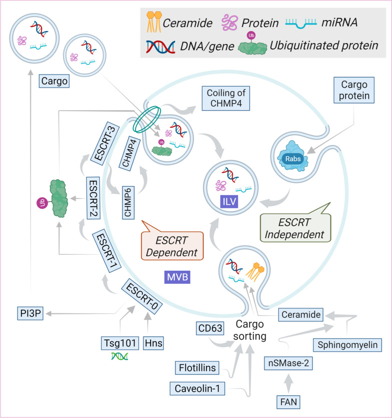
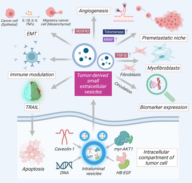
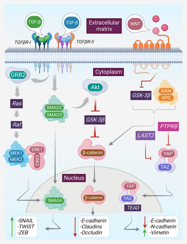
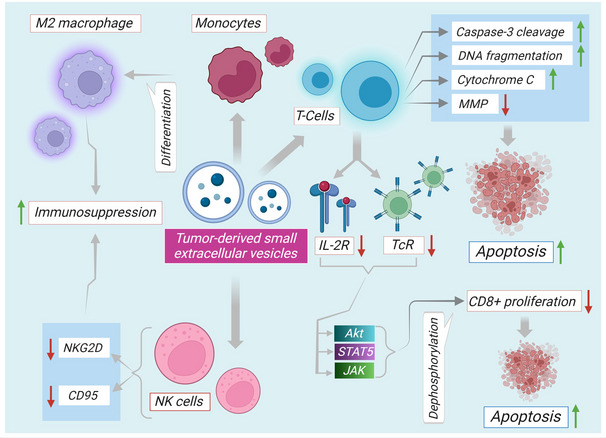
The production and release of tumor-derived small extracellular vesicles (TDSEVs) from cancerous cells play a pivotal role in the propagation of cancer, through genetic and biological communication with healthy cells. TDSEVs are known to orchestrate the invasion-metastasis cascade via diverse pathways. Regulation of early metastasis processes, pre-metastatic niche formation, immune system regulation, angiogenesis initiation, extracellular matrix (ECM) remodeling, immune modulation, and epithelial-mesenchymal transition (EMT) are among the pathways regulated by TDSEVs. MicroRNAs (miRs) carried within TDSEVs play a pivotal role as a double-edged sword and can either promote metastasis or inhibit cancer progression. TDSEVs can serve as excellent markers for early detection of tumors, and tumor metastases. From a therapeutic point of view, the risk of cancer metastasis may be reduced by limiting the production of TDSEVs from tumor cells. On the other hand, TDSEVs represent a promising approach for in vivo delivery of therapeutic cargo to tumor cells. The present review article discusses the recent developments and the current views of TDSEVs in the field of cancer research and clinical applications.

Keywords: Biomarkers; Cancer; Epithelial-mesenchymal transition; Metastasis; MicroRNA; Oncogenic transformation; Tumor microenvironment; Tumor-derived small extracellular vesicles.


Conflict of interest statement

The authors declare no competing interests.

Figures

Fig. 1
Fig. 1
Schematic representation of biogenesis of ILVs. ILVs are the precursors of exosomes created by the inward budding of microdomains and their fission. Assembly of ESCRT machinery starts with the localization of ESCRT-0 on EEs. PI3P aids in recruiting early ESCRT protein Hrs. Hrs then binds with Tsg101 to involve ESCRT-I in the process. ESCRT-I, in turn, binds with ESCRT-II. Subsequent binding of CHMP6 of ESCRT-III with ESCRT-II activates CHMP4 to the endosomal membrane. CHMP4, along with ubiquitinated protein play a pivot in the inward budding of the membrane to form ILVs containing proteins, DNA and miRs. Polymerization of CHMP4 forming spiral coils store potential energy, on elastic compression, this energy gets released giving rise to negative curvature within the membrane As part of the endocytic mechanism, exosome precursors are discharged into MVBs. Also, ESCRT-independent mechanisms involving ceramide, tetraspanins (CD63), and Rabs (Rab31), respectively, have been proposed. In the nSMase2-ceramide pathway, FAN upregulates ceramide production from sphingomyelin. Ceramide, self-associates to form raft structures within the cell membrane to initiate the formation of curvature and subsequent budding. Caveolin-1 and flotilin play vital roles in raft formation and sorting activities. After the sorting procedures are finished, MVBs actively bypass lysosomal fusion. Rabs ensure to prevent MVBs from degradation before fusing with the plasma membrane. Sphingomyelin tends to reorganize the plasma membrane into lipid raft microdomains which subsequently trigger negative curvature in the membrane. Arrows indicate downstream cellular events. EE, early endosome; ESCRT, an endosomal sorting complex required for transport; ILVs, intraluminal vesicles; miR, microRNA; MVB, multivesicular body
Fig. 2
Fig. 2
Pharmacological mechanisms and properties of TDSEVs. Components of TDSEVs participate in intercellular communication and management of tumor microenvironment. TDSEVs contain a plethora of proteins and enzymes engaged in metabolic processes. ILVs are primarily characterized by abnormal production of several different oncoproteins, including caveolin-1, HB-EGF, and MyrAkt1 TDSEVs orchestrate tumorigenic cascade via regulating early metastasis processes, pre-metastatic niche formation carrying telomerase and MMIF, immune system regulation, angiogenesis initiation, ECM remodeling, EMT pathway. TGF-β is transferred by TDSEVs from cancerous cells to healthy fibroblasts, increasing the development of myofibroblasts. DNA within TDSEVs exhibit major translational significance by regulating circulating biomarkers aiding in the early identification of cancer and metastasis. SEV miRs, can in turn lead to a pro-metastatic inflammatory response involving cytokines TNF-α and interleukins. Interestingly, TRAIL, from TDSEVs can restore apoptosis at tumor sites. ‘↑’ represents upregulation. Arrows indicate downstream cellular events/activation
Fig. 3
Fig. 3
Regulation of intracellular and extracellular markers towards EMT. EMT is regulated by intracellular and extracellular markers. Wnt signaling inhibits GSK-3β which is an inhibitor of β-catenin, β-catenin in turn downregulates epithelial marker E-cadherin through influencing transcriptional factors. YAP/TAZ translocates to the nucleus and binds to TEAD to enhance the mesenchymal markers i.e. vimentin and N-cadherin and downregulate E-cadherin. TGF-β signaling can trigger the MAPK/ERK pathway and Smad pathway. Smad inhibits GSK-3β and can activate transcription factors including SNAIL, ZEB, and TWIST, resulting in a loss of cell–cell adhesion and an increase in mesenchymal markers. ERK pathway also contributes to Smad4 at the nucleus in the enhancement of the mesenchymal transcription factors.‘↑’ indicates upregulation, ‘↓’ indicates downregulation. Arrows indicate downstream cellular events/activation and lines indicate inhibition. Akt, Ak strain transforming; EMT, epithelial-mesenchymal transition; GSK-3β, glycogen synthase kinase-3 beta; PTPRB, receptor protein tyrosine phosphatase; TAZ, transcriptional coactivator with PDZ-binding motif; TEAD, transcriptional enhanced associate domain; Wnt, Wingless-related integration site; YAP, yes-associated protein; ZEB, Zinc-finger E-box-binding homeobox
Fig. 4
Fig. 4
Immune modulatory pathways through TDSEVs. TDSEVs exert detrimental effects on TcR and IL-2R activities. TDSEVs can reduce JAK expression and phosphorylation in activated T cells. TDSEVs can also boost CD4 + T cell expression while lowering CD8 + T cell proliferation. TDSEVs, once again, can increase STAT5 phosphorylation in activated CD4 + T cells while decreasing STAT5 phosphorylation in active CD8 + T cells. TDSEV-mediated apoptosis is characterized by DNA fragmentation, caspase-3 cleavage, mitochondrial cytochrome C release, and MMP loss. TDSEVs also impact the PI3K/Akt signaling pathway to increase apoptosis via Akt dephosphorylation. TDSEVs can also excite NK cells by activating NKG2D receptors on the surface of SEVs. Interestingly, cancer cell growth, invasion, and migration can be aided by M2 macrophage ‘↑’ indicates upregulation, ‘↓’ indicates downregulation. Arrows indicate downstream cellular events/activation. IL-2R. interleukin 2 receptor; MMP. Mitochondrial membrane potential TcR, T cell receptor

References

    1. Théry C, Witwer KW, Aikawa E, Alcaraz MJ, Anderson JD, Andriantsitohaina R, et al. Minimal information for studies of extracellular vesicles 2018 (MISEV2018): a position statement of the International Society for Extracellular Vesicles and update of the MISEV2014 guidelines. J Extracell Vesicles. 2018;7(1):1535750. doi: 10.1080/20013078.2018.1535750. - DOI - PMC - PubMed
    1. Spugnini EP, Logozzi M, Di Raimo R, Mizzoni D, Fais S. A Role of Tumor-Released Exosomes in Paracrine Dissemination and Metastasis. Int J Mol Sci. 2018;19(12):3968. doi: 10.3390/ijms19123968. - DOI - PMC - PubMed
    1. Ahmadi M, Rezaie J. Tumor cells derived-exosomes as angiogenenic agents: possible therapeutic implications. J Transl Med. 2020;18(1):249. doi: 10.1186/s12967-020-02426-5. - DOI - PMC - PubMed
    1. Li K, Chen Y, Li A, Tan C, Liu X. Exosomes play roles in sequential processes of tumor metastasis. Int J Cancer. 2019;144(7):1486–1495. doi: 10.1002/ijc.31774. - DOI - PubMed
    1. Vidal M. Exosomes: Revisiting their role as "garbage bags". Traffic. 2019;20(11):815–818. doi: 10.1111/tra.12687. - DOI - PubMed

Publication types