. 2024 Jan 20;26(1):31.
doi: 10.1186/s13075-024-03263-3.

Potential role of RhoA GTPase regulation in type interferon signaling in systemic lupus erythematosus

Affiliations

Potential role of RhoA GTPase regulation in type interferon signaling in systemic lupus erythematosus

Wei Fan et al. Arthritis Res Ther. .

Abstract

Objective: Systemic lupus erythematosus (SLE) is an autoimmune disorder characterized by abnormal activation of the type I interferon (IFN) pathway, which results in tissue inflammation and organ damage. We explored the role of the RhoA GTPase in the type I IFN activation pathway to provide a potential basis for targeting GTPase signaling for the treatment of SLE.

Methods: Total RNA was extracted from peripheral blood mononuclear cells (PBMCs) of SLE patients and healthy controls, and the mRNA expression levels of RhoA and IFN-stimulated genes were measured by SYBR Green quantitative reverse transcriptase-polymerase chain reaction. IFN-a-stimulated response element (ISRE)-luciferase reporter gene assays and Western blotting were conducted to assess the biologic function of RhoA. An enzyme-linked immunoassay (ELISA) measured C-X-C motif chemokine ligand 10 (CXCL10) protein expression.

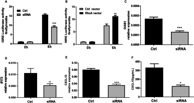
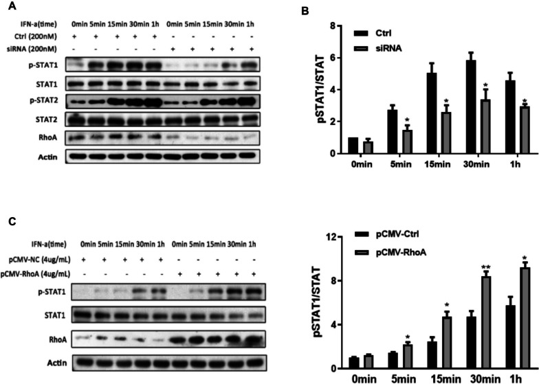
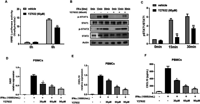
Results: Our studies demonstrate that the expression of RhoA in the PBMCs of SLE subjects was significantly higher than in healthy controls and positively correlated with type I IFN scores and type I IFN-stimulated gene (ISGs) expression levels. SiRNA-mediated knockdown of RhoA and the RhoA/ROCK inhibitor Y27632 reduced the activity of the type I IFN-induced ISRE, the signal transducer and activator of transcription 1 (STAT-1) phosphorylation, and the expression of CXCL10 and 2'-5'-oligoadenylate synthetase 1 (OAS1). Finally, we verified that Y27632 could significantly down-regulate the OAS1 and CXCL10 expression levels in the PBMCs of SLE patients.

Conclusion: Our study shows that RhoA positively regulates the activation of the type I IFN response pathway. Reducing the expression level of RhoA inhibits the abnormal activation of the type I IFN system, and the RhoA/ROCK inhibitor Y27632 decreases aberrant type I IFN signaling in SLE PBMCs, suggesting the possibility of targeting the RhoA GTPase for the treatment of SLE.

Keywords: Autoimmunity; RhoA; RhoA/ROCK inhibitor; Systemic lupus erythematosus; Type I IFN.


Conflict of interest statement

The authors declare no competing interests.

Figures

Fig. 1
Fig. 1
Elevated RhoA expression correlates positively with type I IFN-inducible gene expression and IFN scores. A Significantly higher expression of RhoA mRNA in the PBMCs of SLE patients (n = 36) than in healthy controls (HC, n = 60). BE Correlations between RhoA expression and the type I IFN-inducible genes OAS1, CXCL10, IFIT3, and MX1 in SLE patients. F Type I IFN scores calculated by integrating the relative expression of the OAS1, CXCL10, IFIT3, and MX1 genes in the SLE and HC groups. G RhoA was highly expressed in high-type I IFN-scoring patients. The dotted line represents the mean ± 2SD of the HC values. H Positive correlation between type I IFN scores and SLEDAI values. I Positive correlation between RhoA expression levels and SLEDAI values. Bars in A and G show the mean ± SEM. Each symbol represents an individual sample. Expression is defined as the relative expression of the gene of interest in comparison to GAPDH. *p < 0.05, ***p < 0.001 A and G by Mann–Whitney U test and B-F, H, and I by Spearman rank correlation test
Fig. 2
Fig. 2
RhoA regulates the type I IFN-stimulated response element (ISRE) and downstream gene expression. A ISRE activity of HEK293T cells transfected with ISRE-luciferase and a Renilla reporter plasmid together with a negative control (Ctrl) or RhoA-targeted siRNA (each at 200 nM). B ISRE activity of HEK293T cells transfected with RhoA-overexpression or control plasmids (each at 100 ng). At 24 h after transfection, the cells were left unstimulated (0 h) or stimulated with IFN-a (1000 U/mL, 6 h). Data in A and B are expressed as fold-change based on relative luciferase activities (ratio of firefly luciferase to Renilla luciferase). CE The relative expression of OAS1, IFIT3, and CXCL10 mRNAs were determined by quantitative PCR. F The CXCL10 levels in culture supernatants were determined by enzyme-linked immunosorbent assay. Data were assessed in HEK293T (C, D) and THP1 cells (E, F) at 24 h after transfection of the negative control (Ctrl) or siRNA (200 nM) stimulated cells with IFN-a (1000 U/mL) for 6 h. Bars show the mean ± SEM of 3 independent experiments. *p < 0.05, **p < 0.01 and ***p < 0.001 by Student’s t-test
Fig. 3
Fig. 3
RhoA regulates type I IFN-induced STAT-1 phosphorylation. Western blot analysis (A, C) and densitometry histograms (B, D) of cell lysates of cultured HEK293T cells (5 × 105 cells per well) transfected with siRNA (200 nM) targeting RhoA mRNA (A) or a RhoA expression plasmid vector (4 µg/mL) (C), and their controls (a negative control siRNA or pCMV-NC) stimulated with IFN-a (1000 U/mL) for different times, as shown. The staining density histograms are from three independent experiments and values are expressed as the ratios of phosphorylated STAT-1 protein to total STAT-1. The immunoblot of cells transfected with a negative control siRNA (B) or the pCMV-NC vectors (D) were set at a value of 1. *p < 0.05, **p < 0.01.by Student’s t-test
Fig. 4
Fig. 4
The RhoA/Rock Inhibitor Y27632 downregulates type I IFN signaling. A HEK293T cells were co-transfected with ISRE-luciferase and Renilla reporter plasmids for 24 h, then cultured with or without Y27632 (60 μM, 45 min) before the addition of IFN-a (1000 U/mL, 6 h). Luciferase activity was measured by dual luciferase assay. B PBMCs from healthy controls were cultured in the presence or absence of Y27632 (60 μM, 45 min) and stimulated with IFN-a (1000 U/mL) for different times. Western blots show whole-cell lysates harvested at the indicated times. C Histograms show the ratios of phosphorylated to total STAT-1 at the indicated times. The ratio of pSTAT-1/STAT-1 in the absence of Y27632 and IFN-a at 0 min was set at 1. D Relative expression of OAS1 and E CXCL10 mRNA in PBMCs cultured with Y27632 (0, 30, 60, and 90 μM, for 45 min and stimulated with IFN-a (1000 U/mL) for 6 h. Results are the relative expression levels of OAS1 and CXCL10 mRNA normalized to endogenous GAPDH mRNA levels. F CXCL10 levels in PBMC culture supernatants quantified by ELISA. Bars show the mean ± SEM of 3 individual healthy donors; *p < 0.05, **p < 0.01, A and C versus vehicle by Student’s t-test
Fig. 5
Fig. 5
The RhoA/ROCK inhibitor Y27632 reduces type I IFN signaling in lupus PBMCs. A Relative expression of OAS1 and CXCL10 mRNA in PBMCs obtained from SLE patients (n = 6) and incubated in medium alone or with Y27632 (60 μM, 6 h). B OAS1 and C. CXCL10 mRNA measurements in lysates of lupus PBMCs (n = 6) cultured with or without Y27632 (60 μM, 45 min) and stimulated with IFN-a (1000 U/mL). Results are the relative expression levels of OAS1 and CXCL10 mRNA normalized to endogenous GAPDH mRNA levels. D CXCL10 levels in culture supernatants quantified by ELISA. Bars show the mean ± SEM of 6 individual PBMC samples. *p < 0.05 by Student’s t-test

Update of

Similar articles

Cited by

References

    1. Durcan L, O'Dwyer T, Petri M. Management strategies and future directions for systemic lupus erythematosus in adults. Lancet. 2019;393:2332–2343. doi: 10.1016/S0140-6736(19)30237-5. - DOI - PubMed
    1. Jiang J, Zhao M, Chang C, Wu H, Lu Q. Type I interferons in the pathogenesis and treatment of autoimmune diseases. Clin Rev Allergy Immunol. 2020;59:248–272. doi: 10.1007/s12016-020-08798-2. - DOI - PubMed
    1. Rönnblom L. The importance of the type I interferon system in autoimmunity. Clin Exp Rheumatol. 2016;34:21–24. - PubMed
    1. Gallucci S, Meka S, Gamero AM. Abnormalities of the type I interferon signaling pathway in lupus autoimmunity. Cytokine. 2021;146:155633. doi: 10.1016/j.cyto.2021.155633. - DOI - PMC - PubMed
    1. Chyuan IT, Tzeng HT, Chen JY. Signaling pathways of type I and type III interferons and targeted therapies in systemic lupus erythematosus. Cells. 2019;8:963. doi: 10.3390/cells8090963. - DOI - PMC - PubMed