. 2024 Jan 19;16(1):15.
doi: 10.1186/s13073-023-01275-3.

Non-canonical antigens are the largest fraction of peptides presented by MHC class I in mismatch repair deficient murine colorectal cancer

Affiliations

Non-canonical antigens are the largest fraction of peptides presented by MHC class I in mismatch repair deficient murine colorectal cancer

Giuseppe Rospo et al. Genome Med. .

Abstract

Background: Immunotherapy based on checkpoint inhibitors is highly effective in mismatch repair deficient (MMRd) colorectal cancer (CRC). These tumors carry a high number of mutations, which are predicted to translate into a wide array of neoepitopes; however, a systematic classification of the neoantigen repertoire in MMRd CRC is lacking. Mass spectrometry peptidomics has demonstrated the existence of MHC class I associated peptides (MAPs) originating from non-coding DNA regions. Based on these premises we investigated DNA genomic regions responsible for generating MMRd-induced peptides.

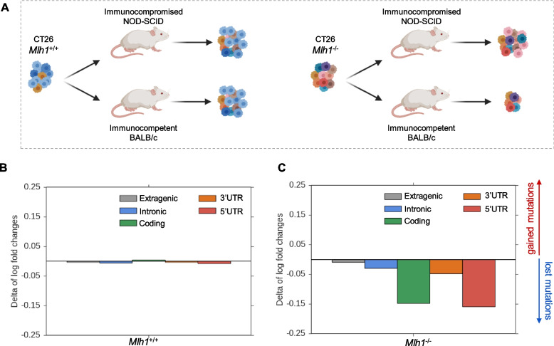
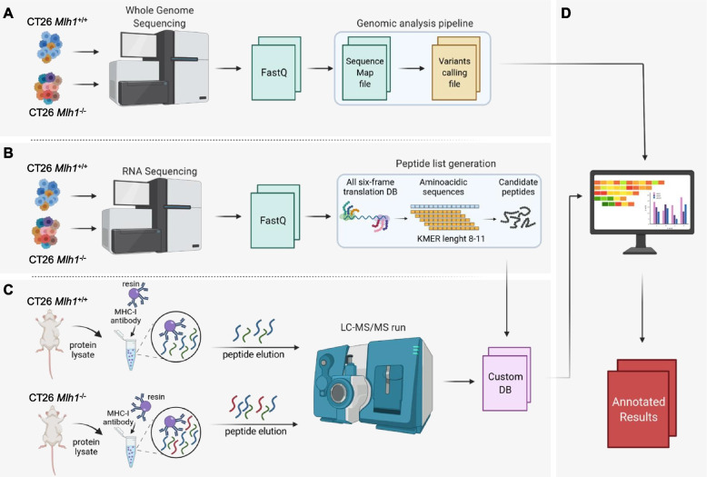
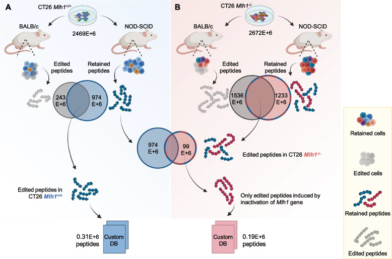
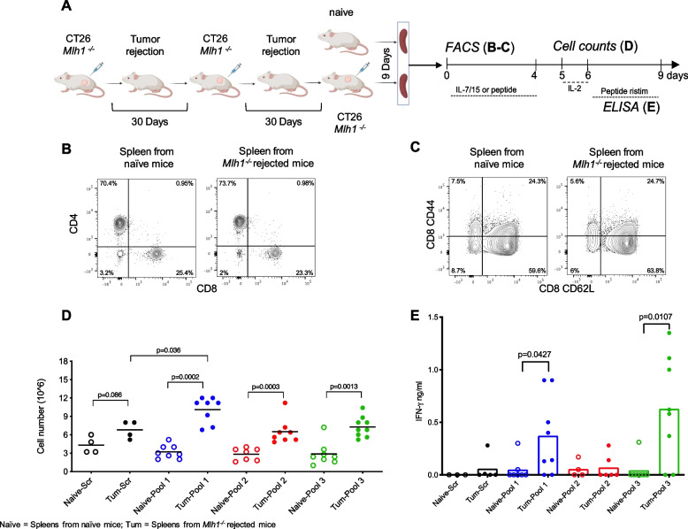
Methods: We exploited mouse CRC models in which the MMR gene Mlh1 was genetically inactivated. Isogenic cell lines CT26 Mlh1+/+ and Mlh1-/- were inoculated in immunocompromised and immunocompetent mice. Whole genome and RNA sequencing data were generated from samples obtained before and after injection in murine hosts. First, peptide databases were built from transcriptomes of isogenic cell lines. We then compiled a database of peptides lost after tumor cells injection in immunocompetent mice, likely due to immune editing. Liquid chromatography-tandem mass spectrometry (LC-MS/MS) and matched next-generation sequencing databases were employed to identify the DNA regions from which the immune-targeted MAPs originated. Finally, we adopted in vitro T cell assays to verify whether MAP-specific T cells were part of the in vivo immune response against Mlh1-/- cells.

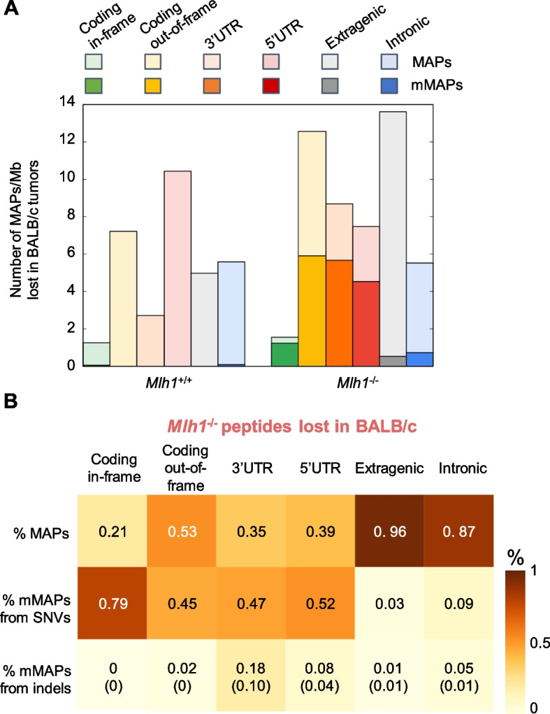
Results: Whole genome sequencing analyses revealed an unbalanced distribution of immune edited alterations across the genome in Mlh1-/- cells grown in immunocompetent mice. Specifically, untranslated (UTR) and coding regions exhibited the largest fraction of mutations leading to highly immunogenic peptides. Moreover, the integrated computational and LC-MS/MS analyses revealed that MAPs originate mainly from atypical translational events in both Mlh1+/+ and Mlh1-/- tumor cells. In addition, mutated MAPs-derived from UTRs and out-of-frame translation of coding regions-were highly enriched in Mlh1-/- cells. The MAPs trigger T-cell activation in mice primed with Mlh1-/- cells.

Conclusions: Our results suggest that-in comparison to MMR proficient CRC-MMRd tumors generate a significantly higher number of non-canonical mutated peptides able to elicit T cell responses. These results reveal the importance of evaluating the diversity of neoepitope repertoire in MMRd tumors.

Keywords: Colorectal cancer; HLA-peptidomics; Immune surveillance; MAPs; Mismatch repair; Neoantigens; Next-generation sequencing; Non-canonical antigens; Non-coding DNA.


Conflict of interest statement

A. Bardelli served in a consulting/advisory role for Illumina and Inivata. A. Bardelli and G.G. are cofounders and shareholders of NeoPhore. A. Bardelli is a member of the NeoPhore scientific advisory board. The remaining authors declare that they do not have any competing interests.

Figures

Fig. 1
Fig. 1
Analysis of mutations targeted in MMR-proficient and MMR-deficient CT26 after injection in immunocompromised and immunocompetent mice. A Experimental workflow employed for the analysis of mutations (SNVs and indels) in WGS data of CT26 Mlh1+/+ and Mlh1-/- samples. Briefly, each CT26 clone was inoculated into NOD-SCID (immunocompromised) and BALB/c (immunocompetent) mice 150 days after genome editing. CT26 MMR-proficient and MMR-deficient tumors underwent WGS at the time of injection and after excision from the mice when tumors reached 1200 mm3 of volume in NOD-SCID mice. In the case of BALB/c mice, the tumors were excised when they reached volumes of 1100 mm3 and 800 mm3 for Mlh1+/+ and Mlh1-/- tumors, respectively. Delta between log fold changes evaluated after injection in immunocompromised and immunocompetent mice in CT26 Mlh1+/+ (B) and CT26 Mlh1-/- (C). Log fold changes analysis of gained and lost alterations was calculated from CT26 Mlh1+/+ and Mlh1-/- pre-injection data, respectively. The alterations were grouped in regions and normalized per Mb before log fold change calculation
Fig. 2
Fig. 2
Development of a pipeline for MAP identification. A WGS data were generated from CT26 Mlh1+/+ and Mlh1-/- samples and analyzed using IDEA pipeline [28] in order to produce the alignment and variant calling files. B RNAseq data were further generated from CT26 Mlh1+/+ and Mlh1-/- cells 150 days after genome editing and after excision from the mice when tumors reached 1200 mm3 of volume in NOD-SCID mice. In the case of BALB/c mice, the tumors were excised when they reached volumes of 1100 mm3 and 800 mm3 for Mlh1+/+ and Mlh1-/- tumors, respectively. FastQ files were handled to produce the list of all putative peptides present in the transcriptome of each sample. In brief, every transcript sequence in the FastQ files underwent all-six frame translation; then the lists of 8–11 amino acid long peptides were generated using the KMER approach; finally, the peptide lists were compared to select only peptides targeted in tumors excised from immunocompetent mice (see methods). C CT26 Mlh1+/+ and Mlh1-/- tumor masses were explanted from NOD-SCID mice (n = 6 per group) and protein extraction was performed. MHC-I was isolated from whole protein lysates through H-2d antibodies conjugated to resin, then peptides were eluted from MHC-I and injected in mass spectrometer. The LC-MS/MS data were then analyzed using MaxQuant. Peptides were searched against the customized DB made of targeted peptides generated by RNAseq data. D Sequence results obtained from the immune-peptidomic pipeline were ultimately matched with WGS data to retrieve information about the genomic sources of targeted peptides (see “Methods”)
Fig. 3
Fig. 3
Identification of targeted MAPs in Mlh1+/+ and Mlh1-/- tumor cells. A The peptide list generated from RNAseq analysis of CT26 Mlh1+/+ cells grown in vitro was compared to the corresponding lists obtained after tumor growth in mice (see Table 2). Thus, peptides lost after injection in CT26 Mlh1+/+ post BALB/c M3 mouse and retrieved after inoculation in CT26 Mlh1+/+ post NOD-SCID M2 mouse were selected. The overlap of these two peptide datasets generated the database of CT26 Mlh1+/+ targeted peptides. B Peptide lists generated from RNAseq analysis in Mlh1-/- samples before and after in vivo growth were compared (see Table 2). This allowed the identification of peptides lost after injection in CT26 Mlh1-/- post BALB/c M2, M6, and M7 mice but maintained in CT26 Mlh1-/- post NOD-SCID M5 mouse. The overlap of these two datasets generated a list of peptides from which specific CT26 Mlh1+/+ sequences were removed. The latter list created the targeted peptides database specific to CT26 Mlh1-/-
Fig. 4
Fig. 4
Characterization of targeted non-canonical MAPs and mMAPs in Mlh1+/+ and Mlh1-/- tumor cells. A The numbers of annotated MAPs were normalized (per Mb) in CT26 Mlh1+/+ and Mlh1-/- samples and are reported in light colors. mMAPs are highlighted in solid colors. B Percentage of mutated and wild-type Mlh1-/- MAPs. Mutated MAPs originated from indels were further labeled with microsatellite information (data in brackets)
Fig. 5
Fig. 5
Non-canonical MAP-specific T cells in mice rejecting Mlh1-/- tumors. A Immunocompetent mice (BALB/c) were injected with 5X10⁵ Mlh1-/- tumor cells per mouse. Upon rejection, mice were re-challenged twice with Mlh1-/- tumor cells (30 days after the previous injection). Nine days after the last injection, mice were sacrificed, and the spleen were surgically resected. Naïve mice were used as control. B Representative events of viable CD4+ and CD8+ T cells are shown in naïve mice and tumor rejected mice. C Single cells were stained with anti-CD4, CD8, CD44, and CD62L mAb and analyzed by FACS. Data depicts naïve and memory markers of viable CD8+T cells. D Splenocytes were cultured with or without synthesized MAP peptides for 4 days. Viable cells were separated on Ficoll gradients and counted. Total counts are depicted. E After an overnight incubation in IL-2, T cells, from naive and tumor-vaccinated mice, were either restimulated or not with the indicated peptide pools. A scramble peptide (Ctrl) served as control. The supernatant was collected after 48 h to quantify IFN-γ. The data show the release of IFN-γ from splenocytes of individual mice pulsed with scramble or peptide pools. The values of IFN-γ from unpulsed splenocytes were subtracted from the values represented in the figure. p-values were calculated by Mann–Whitney non-parametric test

Similar articles

Cited by

References

    1. Jiricny J. The multifaceted mismatch-repair system. Nat Rev Mol Cell Biol. 2006;7(5):335–346. doi: 10.1038/nrm1907. - DOI - PubMed
    1. Germano G, Amirouchene-Angelozzi N, Rospo G, Bardelli A. The clinical impact of the genomic landscape of mismatch repair-deficient cancers. Cancer Discov. 2018;8(12):1518–1528. doi: 10.1158/2159-8290.CD-18-0150. - DOI - PubMed
    1. Richman S. Deficient mismatch repair: read all about it (Review) Int J Oncol. 2015;47(4):1189–1202. doi: 10.3892/ijo.2015.3119. - DOI - PMC - PubMed
    1. Woerner SM, Tosti E, Yuan YP, Kloor M, Bork P, Edelmann W, et al. Detection of coding microsatellite frameshift mutations in DNA mismatch repair-deficient mouse intestinal tumors. Mol Carcinog. 2015;54(11):1376–1386. doi: 10.1002/mc.22213. - DOI - PubMed
    1. Boland CR, Goel A. Microsatellite instability in colorectal cancer. Gastroenterology. 2010;138(6):2073–87.e3. doi: 10.1053/j.gastro.2009.12.064. - DOI - PMC - PubMed

Publication types

Supplementary concepts