

Lymphocyte apoptosis and its association with the inflammatory markers and disease severity in juvenile-onset systemic lupus erythematosus patients
- PMID: 38243322
- PMCID: PMC10799351
- DOI: 10.1186/s12969-024-00953-9
Lymphocyte apoptosis and its association with the inflammatory markers and disease severity in juvenile-onset systemic lupus erythematosus patients
Abstract
Background: The defective clearance of apoptotic bodies in juvenile-onset systemic lupus erythematosus (jSLE) potentially leads to the persistence of autoreactive lymphocytes and the perpetuation of the autoimmune response. These factors contribute to the disturbance in lymphocyte apoptosis and show potential as key determinants in the clinical course and severity of jSLE. This study evaluates the role of peripheral blood (PB) lymphocyte apoptosis in prognosis of jSLE and as a predictor for disease activity.
Methods: The study involved 100 jSLE patients and 50 healthy controls. Flow cytometry was used to analyze percentages of lymphocyte apoptosis in PB of all study participants. Plasma levels of pro-inflammatory cytokines were determined using ELISA.
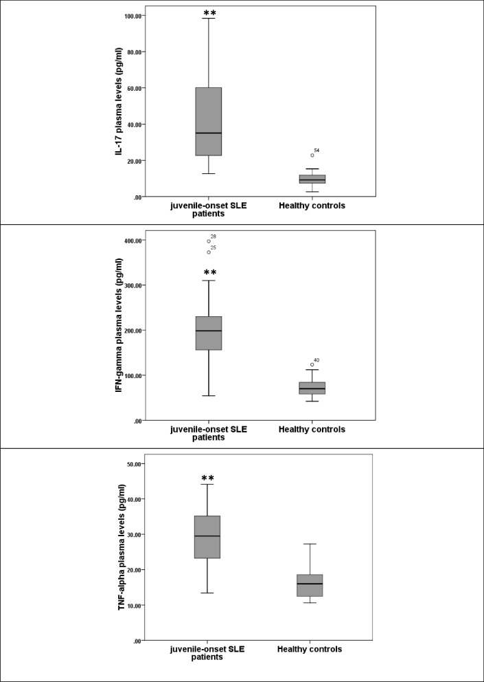
Results: Our results showed that percentages of lymphocyte apoptosis in PB of jSLE patients are significantly higher than those of healthy controls. These percentages are significantly positively associated with disease activity of patients (SLEDAI-2 K). Furthermore, plasma cytokine levels (IL-17, IFN-γ and TNF-α) are significantly elevated in jSLE patients compared to their levels in healthy controls. Also, there are weak significant positive correlations between percentages of PB lymphocyte apoptosis and each of IL-17 and IFN-γ plasma levels in jSLE patients. Moreover, PB lymphocyte apoptosis percentages among jSLE patients are higher in the presence of some clinical and laboratory features than those in their absence.
Conclusion: Peripheral apoptotic lymphocytes could contribute to the prognosis of jSLE and could be used as a predictor for disease activity in jSLE patients.
Keywords: Flow cytometry; IFN-γ; IL-17; Inflammatory cytokines; Juvenile-onset systemic lupus erythematosus; Lymphocyte apoptosis; TNF-α.
© 2024. The Author(s).
Conflict of interest statement
The authors declare that they have no competing interests.
Figures

Similar articles
-
Association of microRNA-125a with the clinical features, disease activity and inflammatory cytokines of juvenile-onset lupus patients.Lupus. 2021 Jun;30(7):1180-1187. doi: 10.1177/09612033211010328. Epub 2021 Apr 17. Lupus. 2021. PMID: 33866896
-
Reduced expressions of apoptosis-related proteins TRAIL, Bcl-2, and TNFR1 in NK cells of juvenile-onset systemic lupus erythematosus patients: relations with disease activity, nephritis, and neuropsychiatric involvement.Front Immunol. 2024 Mar 18;15:1327255. doi: 10.3389/fimmu.2024.1327255. eCollection 2024. Front Immunol. 2024. PMID: 38562920 Free PMC article.
-
Increased Fas and Bcl-2 expression on peripheral blood T and B lymphocytes from juvenile-onset systemic lupus erythematosus, but not from juvenile rheumatoid arthritis and juvenile dermatomyositis.Clin Dev Immunol. 2006 Jun-Dec;13(2-4):283-7. doi: 10.1080/17402520600877786. Clin Dev Immunol. 2006. PMID: 17162369 Free PMC article.
-
Juvenile-onset systemic lupus erythematosus: Update on clinical presentation, pathophysiology and treatment options.Clin Immunol. 2019 Dec;209:108274. doi: 10.1016/j.clim.2019.108274. Epub 2019 Oct 31. Clin Immunol. 2019. PMID: 31678365 Review.
-
Neuropsychiatric involvement in juvenile-onset systemic lupus erythematosus (jSLE).Mol Cell Pediatr. 2023 Aug 9;10(1):5. doi: 10.1186/s40348-023-00161-7. Mol Cell Pediatr. 2023. PMID: 37556020 Free PMC article. Review.
Cited by
-
Association between platelet-to-lymphocyte ratio and immune checkpoint inhibitor-induced thyroid dysfunction.Endocrine. 2025 May;88(2):491-500. doi: 10.1007/s12020-025-04164-4. Epub 2025 Jan 22. Endocrine. 2025. PMID: 39838195
References
MeSH terms
Substances
LinkOut - more resources
Full Text Sources
Medical
