Cardiac magnetic resonance-based layer-specific strain in immune checkpoint inhibitor-associated myocarditis
- PMID: 38243390
- PMCID: PMC10966230
- DOI: 10.1002/ehf2.14664
Cardiac magnetic resonance-based layer-specific strain in immune checkpoint inhibitor-associated myocarditis
Abstract
Aims: To assess the different imaging characteristics between corticosteroid-sensitive (CS) and corticosteroid-refractory (CR) immune checkpoint inhibitor-associated myocarditis (ICIaM) with cardiac magnetic resonance (CMR) and the potential CMR parameters in the early detection of CR ICIaM.
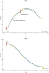
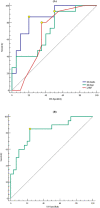
Methods and results: Thirty-five patients diagnosed with ICIaM and 30 age and gender-matched cancer patients without a history of ICI treatment were enrolled. CMR with contrast was performed within 2 days of clinical suspicion. Left ventricular ejection fraction (LVEF) and late gadolinium enhancement (LGE) were assessed by CMR. LV sub-endocardial (GLSendo) and sub-epicardial (GLSepi) global longitudinal strains were quantified by offline feature tracking analysis. CS and CR ICIaM were defined based on the trend of Troponin I and clinical course during corticosteroid treatment. All 35 patients presented with non-fulminant symptoms upon initial assessment. Twenty patients (57.14%) were sensitive, and 15 (42.86%) were refractory to corticosteroids. Compared with controls, 22 patients (62.86%) with ICIaM developed LGE. LVEF decreased in CR ICIaM compared with the CS group and controls. GLSendo (-14.61 ± 2.67 vs. -18.50 ± 2.53, P < 0.001) and GLSepi (-14.75 ± 2.53 vs. -16.68 ± 2.05, P < 0.001) significantly increased in patients with CR ICIaM compared with the CS ICIaM. In patients with CS ICIaM, although GLSepi (-16.68 ± 2.05 vs. -19.31 ± 1.80, P < 0.001) was impaired compared with the controls, GLSendo was preserved. There was no difference in CMR parameters between LGE-positive and negative groups. LVEF, GLSendo, and GLSepi were predictors of CR ICIaM. When LVEF, GLSendo, and GLSepi were included in multivariate analysis, only GLSendo remained an independent predictor of CR ICIaM (OR: 2.170, 95% CI: 1.189-3.962, P = 0.012). A GLSendo of ≥-17.10% (sensitivity, 86.7%; specificity, 80.0%; AUC, 0.860; P < 0.001) could predict CR ICIaM in the ICIaM cohort. Kaplan-Meier analysis showed that in patients with impaired GLSendo of ≥-17.10%, cardiovascular adverse events (CAEs) occurred much earlier than in patients with preserved GLSendo of <-17.10% (Log-rank test P = 0.017).
Conclusions: CR and CS ICIaM demonstrated different functional and morphological characteristics in different myocardial layers. An impaired GLSendo could be a helpful parameter in early identifying corticosteroid-refractory individuals in the ICIaM population.
Keywords: Cardiac magnetic resonance; Global longitudinal strain; Immune checkpoint inhibitor; Late gadolinium enhancement; Myocarditis.
© 2024 The Authors. ESC Heart Failure published by John Wiley & Sons Ltd on behalf of European Society of Cardiology.
Conflict of interest statement
The authors declare that they have no known competing financial interests or personal relationships that could have appeared to influence the work reported in this paper.
Figures


Similar articles
-
The prognostic value of global myocardium strain by CMR-feature tracking in immune checkpoint inhibitor-associated myocarditis.Eur Radiol. 2022 Nov;32(11):7657-7667. doi: 10.1007/s00330-022-08844-x. Epub 2022 May 14. Eur Radiol. 2022. PMID: 35567603
-
Left ventricular myocardial strain and tissue characterization by cardiac magnetic resonance imaging in immune checkpoint inhibitor associated cardiotoxicity.PLoS One. 2021 Feb 19;16(2):e0246764. doi: 10.1371/journal.pone.0246764. eCollection 2021. PLoS One. 2021. PMID: 33606757 Free PMC article.
-
Cardiovascular magnetic resonance in immune checkpoint inhibitor-associated myocarditis.Eur Heart J. 2020 May 7;41(18):1733-1743. doi: 10.1093/eurheartj/ehaa051. Eur Heart J. 2020. PMID: 32112560 Free PMC article.
-
Advances in research on molecular markers in immune checkpoint inhibitor-associated myocarditis.Cancer Innov. 2023 Nov 28;2(6):439-447. doi: 10.1002/cai2.100. eCollection 2023 Dec. Cancer Innov. 2023. PMID: 38125765 Free PMC article. Review.
-
Magnetic Resonance Imaging to Detect Cardiovascular Effects of Cancer Therapy: JACC CardioOncology State-of-the-Art Review.JACC CardioOncol. 2020 Jun 16;2(2):270-292. doi: 10.1016/j.jaccao.2020.04.011. eCollection 2020 Jun. JACC CardioOncol. 2020. PMID: 34396235 Free PMC article. Review.
Cited by
-
Left ventricular global longitudinal strain in patients treated with immune checkpoint inhibitors.Front Oncol. 2024 Dec 24;14:1453721. doi: 10.3389/fonc.2024.1453721. eCollection 2024. Front Oncol. 2024. PMID: 39777349 Free PMC article.
-
Diagnostic and Prognostic Value of Cardiac Magnetic Resonance for Cardiotoxicity Caused by Immune Checkpoint Inhibitors: A Systematic Review and Meta-Analysis.Rev Cardiovasc Med. 2025 Feb 21;26(2):25508. doi: 10.31083/RCM25508. eCollection 2025 Feb. Rev Cardiovasc Med. 2025. PMID: 40026491 Free PMC article.
References
MeSH terms
Substances
Grants and funding
- 82170359/National Natural Science Foundation of China
- SHDC22023207/The Standardized Technology Management and Promotion Project of Shanghai Shenkang Hospital Development Center
- 2020ZSLC21/The Clinical Research Project of Zhongshan Hospital
- 202040344/The Shanghai Municipal Health Commission Fund
- 2020ZSQN74/The Youth Fund of Zhongshan Hospital, Fudan University
LinkOut - more resources
Full Text Sources

