

The role of stromal cells in epithelial-mesenchymal plasticity and its therapeutic potential
- PMID: 38244071
- PMCID: PMC10799841
- DOI: 10.1007/s12672-024-00867-8
The role of stromal cells in epithelial-mesenchymal plasticity and its therapeutic potential
Abstract
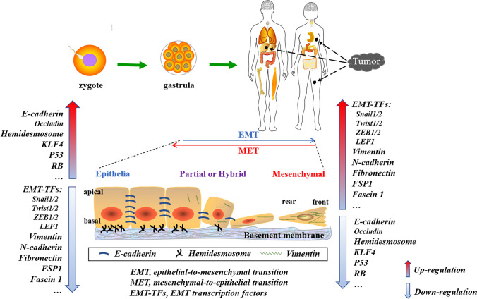
The epithelial-mesenchymal transition (EMT) is a critical tumor invasion and metastasis process. EMT enables tumor cells to migrate, detach from their original location, enter the circulation, circulate within it, and eventually exit from blood arteries to colonize in foreign sites, leading to the development of overt metastases, ultimately resulting in death. EMT is intimately tied to stromal cells around the tumor and is controlled by a range of cytokines secreted by stromal cells. This review summarizes recent research on stromal cell-mediated EMT in tumor invasion and metastasis. We also discuss the effects of various stromal cells on EMT induction and focus on the molecular mechanisms by which several significant stromal cells convert from foes to friends of cancer cells to fuel EMT processes via their secretions in the tumor microenvironment (TME). As a result, a better knowledge of the role of stromal cells in cancer cells' EMT may pave the path to cancer eradication.
Keywords: EMT; Secretions; Stromal cells; Tumor metastasis; Tumor microenvironments.
© 2024. The Author(s).
Conflict of interest statement
All authors declare they have no conflict interest.
Figures


Similar articles
-
Tumor microenvironment and noncoding RNAs as co-drivers of epithelial-mesenchymal transition and cancer metastasis.Dev Dyn. 2018 Mar;247(3):405-431. doi: 10.1002/dvdy.24548. Epub 2017 Sep 18. Dev Dyn. 2018. PMID: 28691356 Review.
-
TGF-β1 dominates stromal fibroblast-mediated EMT via the FAP/VCAN axis in bladder cancer cells.J Transl Med. 2023 Jul 17;21(1):475. doi: 10.1186/s12967-023-04303-3. J Transl Med. 2023. PMID: 37461061 Free PMC article.
-
The dynamic tumor-stromal crosstalk: implications of 'stromal-hot' tumors in the process of epithelial-mesenchymal transition in breast cancer.Mol Biol Rep. 2023 Jun;50(6):5379-5393. doi: 10.1007/s11033-023-08422-4. Epub 2023 Apr 12. Mol Biol Rep. 2023. PMID: 37046108 Review.
-
Tumour microenvironment: a non-negligible driver for epithelial-mesenchymal transition in colorectal cancer.Expert Rev Mol Med. 2021 Nov 11;23:e16. doi: 10.1017/erm.2021.13. Expert Rev Mol Med. 2021. PMID: 34758892 Review.
-
Proteomics analysis reveals the role of ubiquitin specific protease (USP47) in Epithelial to Mesenchymal Transition (EMT) induced by TGFβ2 in breast cells.J Proteomics. 2020 May 15;219:103734. doi: 10.1016/j.jprot.2020.103734. Epub 2020 Mar 19. J Proteomics. 2020. PMID: 32201364
Cited by
-
Single-cell Analysis Highlights Anti-apoptotic Subpopulation Promoting Malignant Progression and Predicting Prognosis in Bladder Cancer.Cancer Inform. 2025 Feb 26;24:11769351251323569. doi: 10.1177/11769351251323569. eCollection 2025. Cancer Inform. 2025. PMID: 40018511 Free PMC article.
-
Factors Determining Epithelial-Mesenchymal Transition in Cancer Progression.Int J Mol Sci. 2024 Aug 17;25(16):8972. doi: 10.3390/ijms25168972. Int J Mol Sci. 2024. PMID: 39201656 Free PMC article. Review.
-
The crosstalk between primary MSCs and cancer cells in 2D and 3D cultures: potential therapeutic strategies and impact on drug resistance.Stem Cells Transl Med. 2024 Dec 16;13(12):1178-1185. doi: 10.1093/stcltm/szae077. Stem Cells Transl Med. 2024. PMID: 39418131 Free PMC article. Review.
References
Publication types
LinkOut - more resources
Full Text Sources
Research Materials