Review
. 2024 Feb;161(2):99-132.
doi: 10.1007/s00418-023-02259-5. Epub 2024 Jan 20.

The peroxisome: an update on mysteries 3.0

Affiliations
Review

The peroxisome: an update on mysteries 3.0

Rechal Kumar et al. Histochem Cell Biol. 2024 Feb.

Abstract

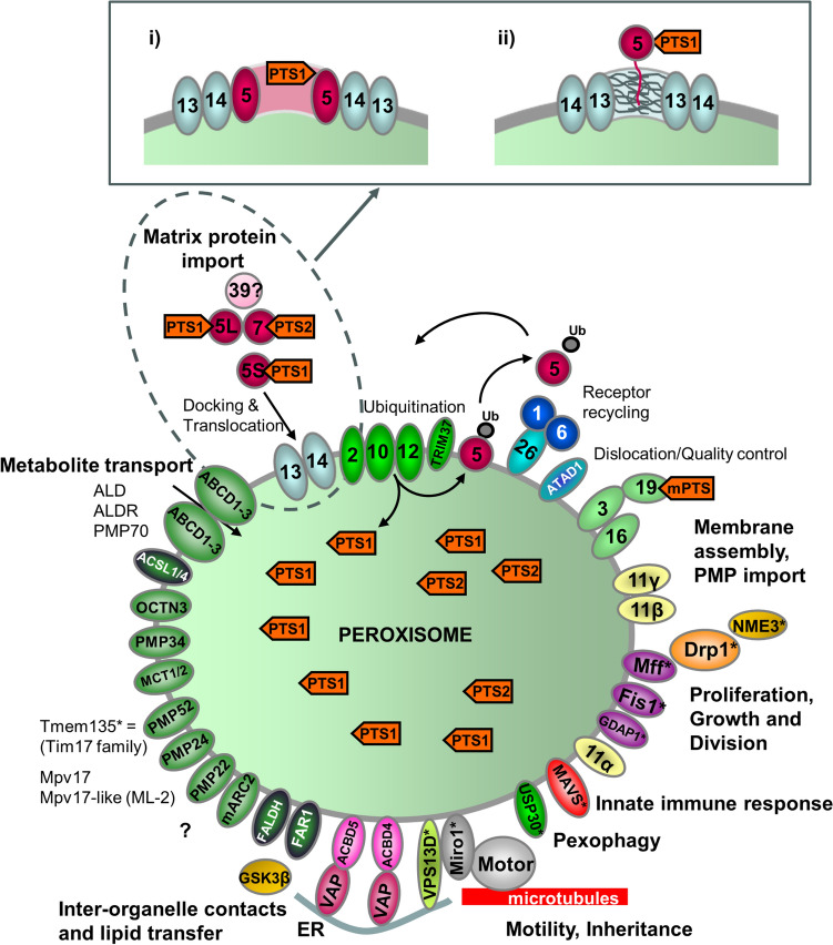
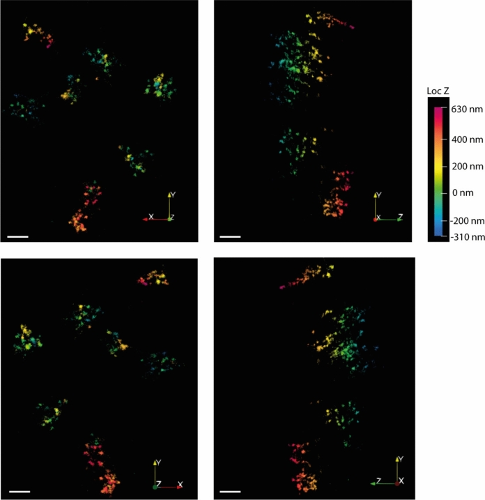
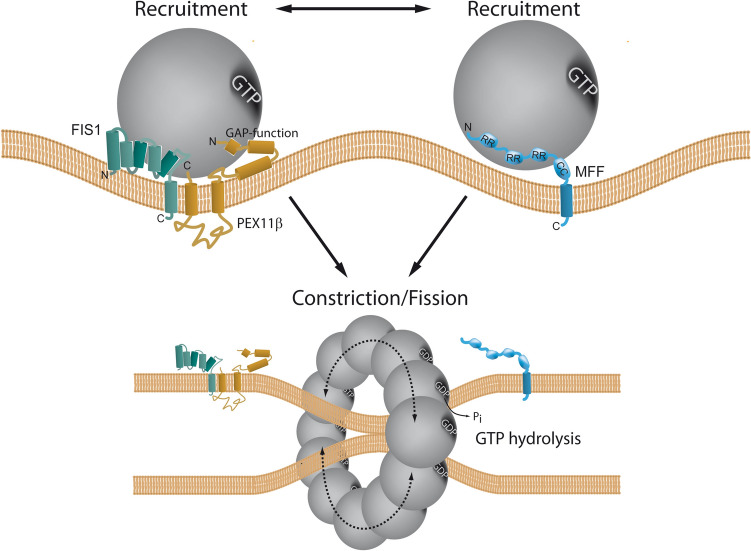
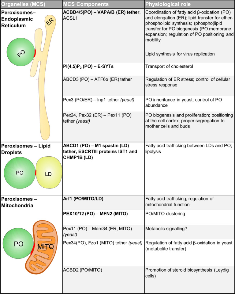
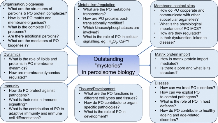
Peroxisomes are highly dynamic, oxidative organelles with key metabolic functions in cellular lipid metabolism, such as the β-oxidation of fatty acids and the synthesis of myelin sheath lipids, as well as the regulation of cellular redox balance. Loss of peroxisomal functions causes severe metabolic disorders in humans. Furthermore, peroxisomes also fulfil protective roles in pathogen and viral defence and immunity, highlighting their wider significance in human health and disease. This has sparked increasing interest in peroxisome biology and their physiological functions. This review presents an update and a continuation of three previous review articles addressing the unsolved mysteries of this remarkable organelle. We continue to highlight recent discoveries, advancements, and trends in peroxisome research, and address novel findings on the metabolic functions of peroxisomes, their biogenesis, protein import, membrane dynamics and division, as well as on peroxisome-organelle membrane contact sites and organelle cooperation. Furthermore, recent insights into peroxisome organisation through super-resolution microscopy are discussed. Finally, we address new roles for peroxisomes in immune and defence mechanisms and in human disorders, and for peroxisomal functions in different cell/tissue types, in particular their contribution to organ-specific pathologies.

Keywords: Membrane contact sites; Motility; Organelle biogenesis; Organelle division; Organelle dynamics; Peroxin; Peroxisome; Protein import; STED microscopy.


Conflict of interest statement

The authors declare no conflict of interest.

Figures

Fig. 1
Fig. 1
Schematic overview of the molecular machineries and proteins localised at the membranes of mammalian peroxisomes. Adapted from Schrader and Fahimi (2008). Related overviews for yeast and zebrafish peroxisomes can be found in Islinger et al. (2018) and Kamoshita et al. (2022). See text for further details. Matrix protein import: After synthesis on free ribosomes, cargo proteins containing the peroxisomal targeting signals PTS1 or PTS2 bind to the corresponding cytosolic receptors PEX5 or PEX7 and form receptor-cargo complexes. The PEX7–cargo complex requires accessory factors for import (PEX5L, a long isoform of PEX5, in mammals and plants, Pex18p and Pex21p in Saccharomyces cerevisiae, Pex20p in Neurospora crassa, Yarrowia lipolytica, and Hansenula polymorpha) (see Table 1). The newly discovered PEX39 may be involved in PTS2-mediated protein import. Import is achieved by a complex set of integral or peripheral PMPs that form the matrix protein import machinery, which mediates docking of the cargo-bound import receptor at the peroxisomal membrane, cargo translocation into the matrix of the organelle by a dynamic translocon, and export of the receptor back to the cytosol. Different models (top) have been proposed (see text for details): (i) PEX5 inserts into the peroxisomal membrane to form an oligomeric transient pore; (ii) PEX13 forms a stable pore, with YG repeats creating a hydrogel, with which PEX5 interacts. Recycling of the receptor involves its ubiquitination (Ub) and extraction from/through the membrane by an AAA-ATPase complex (PEX1, PEX6). PEX6 binds to PEX26 (Pex15p in yeast; see Table 1). Membrane assembly and insertion of PMPs (containing an mPTS) depends on PEX19, PEX3, and PEX16. PEX19 functions as a cycling receptor/chaperone, which binds the PMPs in the cytosol and interacts with PEX3 at the peroxisomal membrane. Proliferation, growth and division: PEX11α, PEX11β, and PEX11γ are involved in the regulation of peroxisome size and number (proliferation) in mammals. Mammalian PEX11β remodels the peroxisomal membrane and interacts with the membrane adaptors MFF and FIS1, which recruit the dynamin-like fission GTPase DRP1 (DRP3A in plants, Vps1p, Dnm1p in S. cerevisiae) to peroxisomes, which is activated by PEX11β. NME3 is supposed to supply GTP for DRP1-mediated fission. Motility: Mammalian peroxisomes move along microtubules, and MIRO1 serves as membrane adaptor for the microtubule-dependent motor proteins kinesin and dynein. Tethering and lipid transfer: ACBD5 and ACBD4 interact with ER-resident VAPA/B to mediate peroxisome-ER contacts in mammals. GSK3β regulates ACBD5-VAP interaction (Kors et al. 2022). VPS13D may be involved in phospholipid transfer from the ER to peroxisomes. ABCD1 is involved in peroxisome-LD contacts via M1 spastin. Metabolite transport: uptake of fatty acids is mediated by ABC transporter proteins (ABCD1-3 in mammals) (ALD, adrenoleukodystrophy protein; ALDR, ALD-related protein). Other transporter and membrane proteins/enzymes: OCTN3, organic cation/carnitine transporter 3; MCT1/2, monocarboxylate transporter 1/2; PMP52 (Tmem135) and PMP24 (PxmP4) belong to the Tim17 family (Žárský and Doležal 2016); members of the PMP22 family are Mpv17, Mpv17-like (ML-P); ACSL1/4, Acyl-CoA synthetase long-chain family member 1/4; Ant1, peroxisomal adenine nucleotide transporter 1; mARC2 (Mosc2), mitochondrial amidoxime reducing component 2; ATAD1/Msp1, ATPase family AAA (ATPase associated with various cellular activities) domain-containing protein 1; FALDH, fatty aldehyde dehydrogenase (Costello et al. 2017a); FAR1, fatty Acyl-CoA reductase 1 (ether lipid biosynthesis); GDAP1, ganglioside-induced differentiation-associated protein 1; MAVS, mitochondrial antiviral signalling protein; TRIM37, tripartite motif-containing protein 37; USP30, ubiquitin-specific protease 30 (Marcassa et al. 2018). Proteins with a dual localization to both peroxisomes and mitochondria are marked with an asterisk. PEX peroxin; PMP peroxisomal membrane protein
Fig. 2
Fig. 2
MINFLUX microscopy of peroxisomes. HEK293 cells expressing the peroxisomal membrane protein PEX26 with a SNAP Tag (SNAP-PEX26), which is labelled with Alexa 647 and imaged on a MINFLUX setup. Each cluster of puncta reflects subdomains of PEX26 in the membrane of a single peroxisome. The figure shows two-dimensional projections of a 3D MINFLUX recording from different angles, as indicated by the coordinates in the lower right corner. The colour code indicates the z position. Scale bar, 200 nm. Figure kindly supplied by K Reglinski/C Eggeling, University of Jena, Germany
Fig. 3
Fig. 3
Schematic of the two alternative pathways of peroxisome division, the PEX11β-FIS1-dependent and the MFF-dependent peroxisome division pathway. CC coiled-coil region; GAP GTPase-activating protein–function; RR repetitive region. Taken from Schrader et al. (2022)
Fig. 4
Fig. 4
Known components of peroxisome-organelle MCSs. For details see text and Silva et al. (2020). ABCD1/3 ATP-binding cassette sub-family D member 1/3; ACBD2/4/5 acyl-coenzyme binding domain protein 2/4/5; ACSL1 long chain fatty-acid-CoA ligase 1; Arf1 ADP-ribosylation factor 1; ATF6α activating transcription factor 6α; CHMP1B charged multivesicular body protein 1B; ER endoplasmic reticulum; ESCRTIII endosomal sorting complexes required for transport III; Fzo1 fuzzy onions homolog 1; Inp1 inheritance of peroxisomes protein 1; IST1 vacuolar protein sorting-associated protein 1; LD lipid droplet; Mdm34 mitochondrial distribution and morphology protein 34; MFN2 mitofusin 2; MITO mitochondrion; M1 spastin, isoform M1 of the microtubule-severing protein spastin; Pex peroxin; PI(4,5)P2 phosphatidylinositol-4,5-biphosphate; PO peroxisome; E-Syts ER-resident extended synaptotagmins; VAPA/B vesicle-associated membrane protein (VAMP)-associated protein A/B. MCS proteins are mammalian unless otherwise stated, and those in bold are addressed further in the text. Adapted from Silva et al. (2020)
Fig. 5
Fig. 5
Overview of some “mysteries” of peroxisome biology yet to be solved. For details see text. PO peroxisome

References

    1. Abe S, Nagai T, Masukawa M, et al. Localization of protein kinase NDR2 to peroxisomes and its role in ciliogenesis. J Biol Chem. 2017;292:4089–4098. doi: 10.1074/jbc.M117.775916. - DOI - PMC - PubMed
    1. Abe Y, Wanders RJA, Waterham HR, et al. Genetic defects in peroxisome morphogenesis (Pex11β, dynamin-like protein 1, and nucleoside diphosphate kinase 3) affect docosahexaenoic acid-phospholipid metabolism. J Inherit Metab Dis. 2023;46:273–285. doi: 10.1002/jimd.12582. - DOI - PubMed
    1. Agbaga MP, Merriman DK, Brush RS, et al. Differential composition of DHA and very-long-chain PUFAs in rod and cone photoreceptors. J Lipid Res. 2018;59:1586–1596. doi: 10.1194/jlr.M082495. - DOI - PMC - PubMed
    1. Albersen M, van der Beek SL, Dijkstra IME, et al. Sex-specific newborn screening for X-linked adrenoleukodystrophy. J Inherit Metab Dis. 2023;46:116–128. doi: 10.1002/jimd.12571. - DOI - PMC - PubMed
    1. Alencastre IS, Rodrigues TA, Grou CP, et al. Mapping the cargo protein membrane translocation step into the PEX5 cycling pathway. J Biol Chem. 2009;284:27243–27251. doi: 10.1074/jbc.M109.032565. - DOI - PMC - PubMed

LinkOut - more resources