

Material Analysis of Explanted Calcified Silicone Intraocular Lenses in Association with Asteroid Hyalosis
- PMID: 38244181
- PMCID: PMC10853093
- DOI: 10.1007/s40123-023-00872-0
Material Analysis of Explanted Calcified Silicone Intraocular Lenses in Association with Asteroid Hyalosis
Abstract
Introduction: The aim of this study was to analyze posterior surface opacification in explanted silicone intraocular lenses (IOLs) with clinicopathologic correlation to asteroid hyalosis.
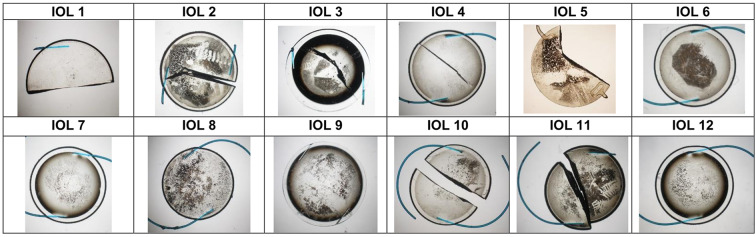
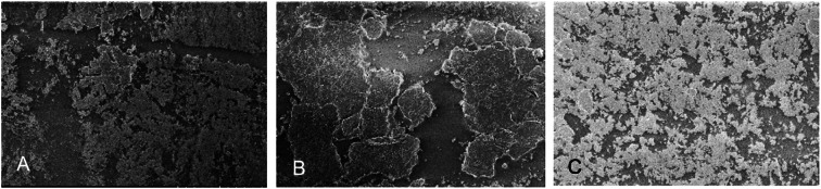
Methods: In a laboratory setup, 12 explanted silicone IOLs underwent laboratory analyses, including light microscopy, scanning electron microscopy (SEM), and energy-dispersive X-ray spectroscopy for elemental composition (EDX). Relevant clinical data were obtained for each case, including gender, age at IOL implantation, dates of implantation and explantation, as well as history of neodymium-dopped yttrium aluminum garnet (Nd:YAG) laser treatments or other opacification removal attempts. High-resolution optical coherence tomography (OCT) images were obtained in vitro with an anterior segment OCT device (Anterion, Heidelberg Engineering, Heidelberg, Germany).
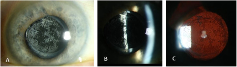
Results: Calcification located at the posterior optic surface of each lens was identified through SEM and EDX analyses, revealing deposits composed of hydroxyapatite. In all cases, IOL polishing using Nd:YAG laser had been attempted prior to IOL exchange. The clinical functional data showed that this type of IOL opacity led to increase in straylight and subjective symptoms of glare.
Conclusions: Silicone IOLs can develop posterior surface calcification in eyes with asteroid hyalosis. There are mechanical techniques of cleaning the IOL surface but in many cases, IOL explantation is the only sustainable way to reduce the patients' straylight levels and glare symptoms. Due to the risk of posterior surface calcification, silicone IOL implantation should be avoided in eyes with asteroid hyalosis.
Keywords: Asteroid hyalosis; IOL calcification; IOL opacification; IOL pathology; Silicone IOLs.
© 2024. The Author(s).
Conflict of interest statement
Lizaveta Chychko, Sonja K. Schickhardt, Hyeck-Soo Son and Ingo Lieberwirth have nothing to disclose. Timur M. Yildirim reports personal fees from Alcon and Hoya and non-financial support from Johnson & Johnson. Gerd U. Auffarth reports grants, personal fees, non-financial support and consulting fees from Johnson & Johnson and Alcon, grants, personal fees and non-financial support from Carl Zeiss Meditec, Hoya, Kowa, Oculentis/Teleon, Rayner, Santen, SIFI, URSAPHARM, grants and personal fees from Biotech, Oculus, EyeYon grants from AcuFocus, Anew, Contamac, Glaukos, Physiol, RHEACELL, outside the submitted work. Ramin Khoramnia reports research grants and lecture fees from Alcon, Hoya, Physiol, Rayner, 1stQ and Johnson & Johnson, lecture fees from Kowa, Ophtec, Teleon, Santen, AcuFocus, Bausch + Lomb and travel grants from Alcon, Teleon, Johnson & Johnson, Rayner and 1stQ outside the submitted work. No funding or sponsorship was received for this study or publication of this article.
Figures

Similar articles
-
Calcification of different designs of silicone intraocular lenses in eyes with asteroid hyalosis.Ophthalmology. 2010 Aug;117(8):1486-92. doi: 10.1016/j.ophtha.2009.12.032. Ophthalmology. 2010. PMID: 20537395
-
Surface calcification of a 3-piece silicone intraocular lens in a patient with asteroid hyalosis: a clinicopathologic case report.Ophthalmology. 2005 Mar;112(3):447-52. doi: 10.1016/j.ophtha.2004.10.025. Ophthalmology. 2005. PMID: 15745772
-
Laser treatment of silicone intraocular lens opacification associated with asteroid hyalosis.Taiwan J Ophthalmol. 2019 Jan-Mar;9(1):49-52. doi: 10.4103/tjo.tjo_65_18. Taiwan J Ophthalmol. 2019. PMID: 30993069 Free PMC article.
-
Complications of cataract and refractive surgery: a clinicopathological documentation.Trans Am Ophthalmol Soc. 2001;99:95-107; discussion 107-9. Trans Am Ophthalmol Soc. 2001. PMID: 11797325 Free PMC article. Review.
-
Akreos Adapt AO Intraocular lens opacification after vitrectomy in a diabetic patient: a case report and review of the literature.BMC Ophthalmol. 2016 Jun 8;16:82. doi: 10.1186/s12886-016-0268-3. BMC Ophthalmol. 2016. PMID: 27277708 Free PMC article. Review.
Cited by
-
Examining Penetration and Residual Depth in Modern Acrylic Foldable Intraocular Lenses: A Laboratory Study Using Differential Interference Contrast Microscopy to Compare Hydrophilic and Hydrophobic Materials.Cureus. 2024 Sep 28;16(9):e70383. doi: 10.7759/cureus.70383. eCollection 2024 Sep. Cureus. 2024. PMID: 39345802 Free PMC article.
References
-
- Łabuz G, Yildirim TM, van den Berg T, Khoramnia R, Auffarth GU. Assessment of straylight and the modulation transfer function of intraocular lenses with centrally localized opacification associated with the intraocular injection of gas. J Cataract Refract Surg. 2018;44:615–622. doi: 10.1016/j.jcrs.2018.01.033. - DOI - PubMed
LinkOut - more resources
Full Text Sources
