. 2024 Mar 6;14(3):jkae012.
doi: 10.1093/g3journal/jkae012.

Expression and potential regulatory functions of Drosophila octopamine receptors in the female reproductive tract

Affiliations

Expression and potential regulatory functions of Drosophila octopamine receptors in the female reproductive tract

Ethan W Rohrbach et al. G3 (Bethesda). .

Abstract

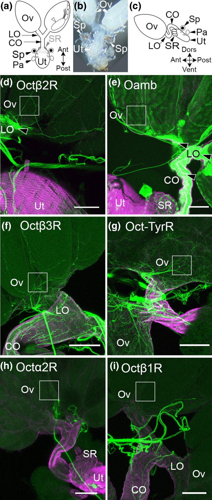
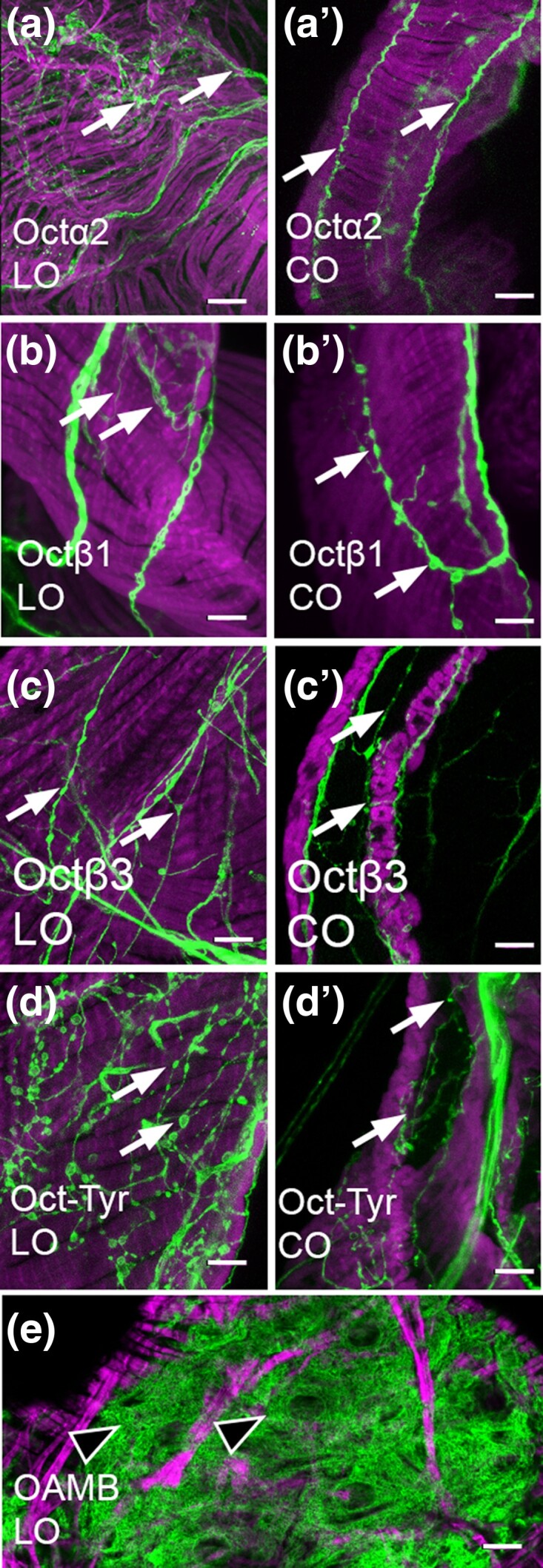
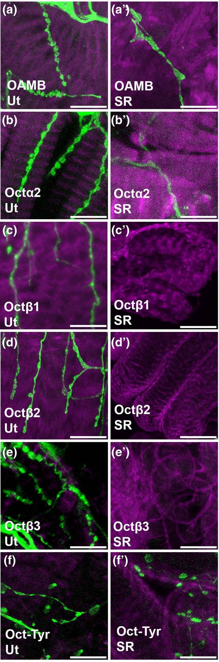
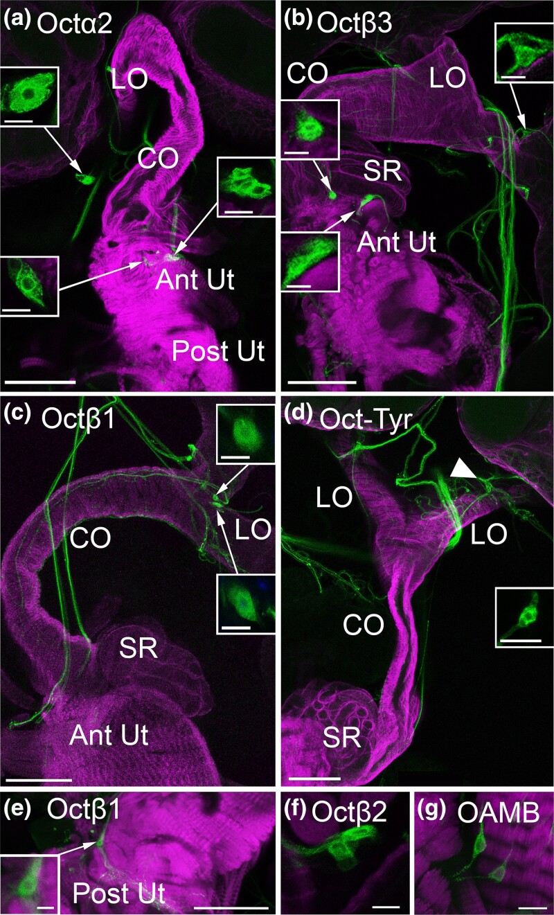
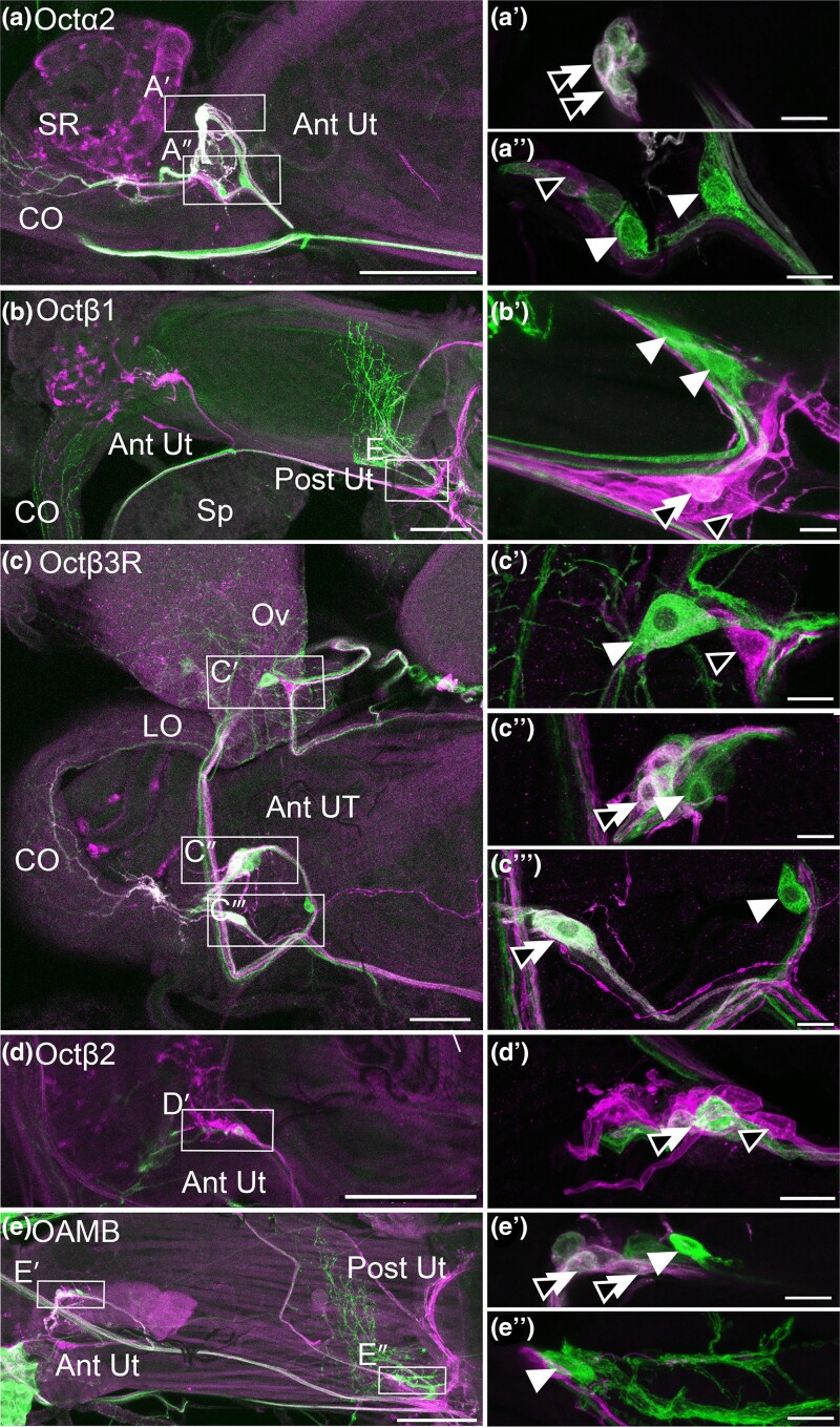
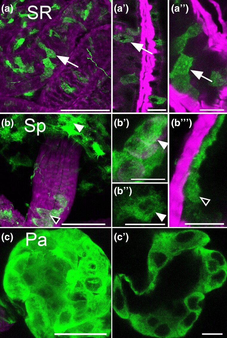
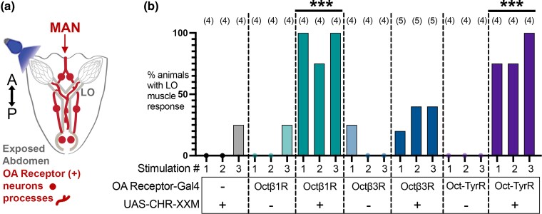
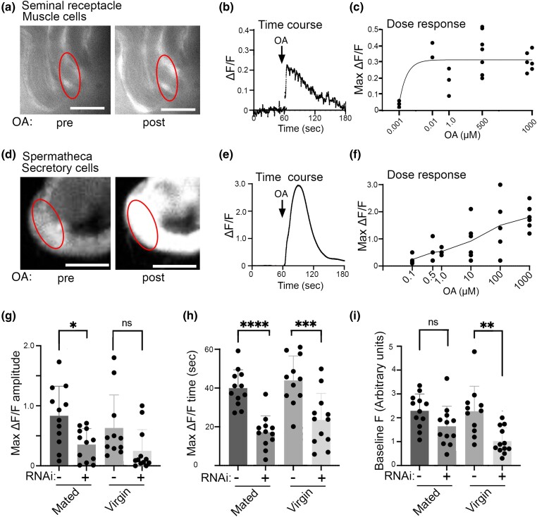
Aminergic signaling is known to play a critical role in regulating female reproductive processes in both mammals and insects. In Drosophila, the ortholog of noradrenaline, octopamine, is required for ovulation as well as several other female reproductive processes. Two octopamine receptors have already been shown to be expressed in the Drosophila reproductive tract and to be required for egg-laying: OAMB and Octβ2R. The Drosophila genome contains 4 additional octopamine receptors-Octα2R, Octβ1R, Octβ3R, and Oct-TyrR-but their cellular patterns of expression in the reproductive tract and potential contribution(s) to egg-laying are not known. In addition, the mechanisms by which OAMB and Octβ2R regulate reproduction are incompletely understood. Using a panel of MiMIC Gal4 lines, we show that Octα2R, Octβ1R, Octβ3R, and Oct-TyrR receptors are not detectable in either epithelium or muscle but are clearly expressed in neurons within the female fly reproductive tract. Optogenetic activation of neurons that express at least 3 types of octopamine receptors stimulates contractions in the lateral oviduct. We also find that octopamine stimulates calcium transients in the sperm storage organs and that its effects in spermathecal, secretory cells, can be blocked by knock-down of OAMB. These data extend our understanding of the pathways by which octopamine regulates egg-laying in Drosophila and raise the possibility that multiple octopamine receptor subtypes could play a role in this process.

Keywords: egg-laying; octopamine; octopamine receptor; oviposition; spermatheca.


Conflict of interest statement

Conflicts of interest The authors declare no conflicts of interest.

Figures

Fig. 1.
Fig. 1.
OA receptor expression in the ovaries. a) A schematized dorsal view of the female Drosophila reproductive tract showing a mature egg (white arrowhead), the ovaries (Ov), lateral oviducts (LO), common oviduct (CO), and the uterus (Ut). Two sperm storage organs are attached to the uterus: the seminal receptacle (SR, indicated in gray because it is on the ventral side of the Ut) and the 2 spermathecae (Sp). An additional pair of parovarian/accessory glands (Pa) is attached to the uterus posterior to the spermatheca. b) A dissected preparation viewed from the dorsal side showing the relative sizes and positions of the Ov, Sp, and Ut. c) A schematize sagittal view of the reproductive tract as it appears in vivo. d–i) MiMIC Gal4 lines for the indicated receptors were used to express membrane-attached GFP (green) and colabeled for phalloidin (magenta) as a marker for muscle cells. The boxed areas highlight the presence or absence of fine processes that ascend into the ovaries. Faint labeling of follicle cells within the boxed area can be seen with OAMB (e). Labeling of the oviduct epithelium is also visible for Octβ2 and OAMB (black arrowheads). Scale bars: 100 μm.
Fig. 2.
Fig. 2.
Processes in the oviducts. a–e). MiMIC Gal4 lines for the indicated receptors were used to express membrane-attached GFP (green) and colabeled for phalloidin (magenta) as in Fig. 1. Fine processes (white arrows) are present for Octα2R, Octβ1R, Octβ3R, and Oct-TyrR as previously reported for Octβ2R and OAMB. Labeling in muscle or epithelium was not detected. An example of epithelial cells expressing OAMB is shown (e, black arrowheads). Scale bars: 10 μm.
Fig. 3.
Fig. 3.
Processes in the uterus and seminal receptacle. a–f) MiMIC Gal4 lines for the indicated receptors were used to express membrane-attached GFP (green) and colabeled for phalloidin (magenta) as in Figs. 1 and 2. Processes in the uterus (a–f) are visible for all receptors. Processes in the seminal receptable (a′–f′) are visible for OAMB, Octα2R, and Oct-TyrR but not Octβ1R, Octβ2R, or Octβ3R. Scale bars: 10 μm.
Fig. 4.
Fig. 4.
Peripheral cell bodies in the reproductive tract. a–g) MiMIC Gal4 lines for the indicated receptors were used to express GFP (green) and colabeled for phalloidin (magenta) as in Figs. 1–3. Cell bodies in the anterior uterus (Ant Ut) express Octα2R (a) and Octβ3R (b), and at least 1 cell body expressing Octβ1R is present in the posterior uterus (e). All other cell bodies shown here that express Octα2R, Octβ3R, Octβ1R, and Oct-TyrR are contained within or attached to free-hanging nerves that connect the MAN to the reproductive tract or connect the anterior and posterior regions of the reproductive tract (a–d). Previously reported cells expressing Octβ2R in the anterior uterus (f) and OAMB in the posterior uterus are shown for comparison (g). Boxed insets show the indicated cells at a higher magnification. Scale bars: (a–e) 100 μm; (f and g and boxed insets in a–d) 10 μm.
Fig. 5.
Fig. 5.
A subset of peripheral cell bodies expressing OA receptors colabel with the marker ppk1.0-LexA. a–e) MiMIC Gal4 lines for the indicated receptors were used to express GFP (green) and colabeled with ppk1.0-LexA expressing RFP (magenta). The boxed regions indicated in a–e are shown at a higher magnification in a′–e′ and a″−e″. Cells labeled for the receptor alone (white arrowhead), ppk1.0 alone (black arrowhead), or both the receptor and ppk1.0 (double white + black arrowheads) are indicated. Scale bars: (a–e) 100 μm; (a′–e′, a″−e″) 10 μm.
Fig. 6.
Fig. 6.
OAMB is expressed in the seminal receptacle and spermathecae. a–c) OAMB(+) cells were marked with GFP and colabeled with phalloidin (magenta). Cells in the seminal receptacle (a–a″, SR, white arrows), the spermathecae (b–b′′′, Sp), and the parovarion glands (c–c″, Pa) express OAMB. In the spermathecae (b–b′′′), cells were labeled in both the bulb (white arrowheads) and the lumen of the stalk (black arrowheads). Scale bars: (a–c) 50 μm; (a–a″, b–b′′′, and c′) 10 μm.
Fig. 7.
Fig. 7.
Optogenetic stimulation of OA receptor–expressing cells drives lateral oviduct contractions. a) Fly abdomens were severed from the throrax, dissected to expose the reproductive organs, and optogenetically stimulated (b). The number of LO contractions in response to each of 3 successive stimulations per fly was quantified. The number of flies per genotype is indicated in parentheses (n). The data were analyzed using Dunnett's test to compare multiple conditions to the negative control UAS-ChRXXM without a Gal4 driver. The control differed significantly (***P < 0.001) from flies expressing UAS-ChRXXM with Octβ1R-Gal4 or Oct-TyrR-Gal4 but not Octβ3R-Gal4 or flies expressing a Gal4 driver in the absence of UAS-ChRXXM.
Fig. 8.
Fig. 8.
OA induces calcium transients in sperm storage organs. UAS-RCaMP1b was expressed in either muscle cells in the seminal receptable with 24B-Gal4 (a–c) or in spermathecal secretory cells with 40B09-Gal4 (d–i) followed by application of OA or vehicle. The red ovals represent examples of ROIs used for quantitation. Sample images (a) of seminal receptacle muscle cells pre- and postaddition (maximal response) of 1 μM OA at 60 s and a trace showing the time course of the response (b). A dose response plot shows seminal receptacle muscle cells increase intracellular calcium in response to OA doses ranging from 0.01 to 1000 μM (c). Sample images (d) of spermathecal secretory cells pre- and postaddition of 1 μM OA at 60 s and the time course of the response (e). Spermathecal secretory cells increase intracellular calcium in response to OA doses ranging from 0.5 to 1000 μM (f). Knocking down OAMB in the spermathecal secretory cells with UAS-OAMB-RNAi (indicated as “RNAi: +”) reduces the maximal RCaMP response to OA in the spermathecal secretory cells of mated flies (g), the time to maximal ΔF/F response in both virgin and mated flies (h), and the baseline RCaMP signal in virgin flies (i, arbitrary units × 103) relative to matched controls (controls express 40B09-Gal4, UAS- RCaMP1b, and UAS-dicer2 but not UAS-OAMB-RNAi. Mean+/− SEM shown; 1-way ANOVA with multiple comparison test across all conditions within each panel, *P ≤ 0.05, **P ≤ 0.01, ***P ≤ 0.001, ****P ≤ 0.0001. Scale bars = 50 μm.

Similar articles

Cited by

References

    1. Allen AK, Spradling AC. 2008. The Sf1-related nuclear hormone receptor Hr39 regulates Drosophila female reproductive tract development and function. Development. 135(2):311–321. doi:10.1242/dev.015156. - DOI - PubMed
    1. Andreatta G, Kyriacou CP, Flatt T, Costa R. 2018. Aminergic signaling controls ovarian dormancy in Drosophila. Sci Rep. 8(1):2030. doi:10.1038/s41598-018-20407-z. - DOI - PMC - PubMed
    1. Avila FW, Bloch Qazi MC, Rubinstein CD, Wolfner MF. 2012. A requirement for the neuromodulators octopamine and tyramine in Drosophila melanogaster female sperm storage. Proc Natl Acad Sci U S A. 109(12):4562–4567. doi:10.1073/pnas.1117689109. - DOI - PMC - PubMed
    1. Balfanz S, Strunker T, Frings S, Baumann A. 2005. A family of octopamine [corrected] receptors that specifically induce cyclic AMP production or Ca2+ release in Drosophila melanogaster. J Neurochem. 93(2):440–451. doi:10.1111/j.1471-4159.2005.03034.x. - DOI - PubMed
    1. Chapman T, Bangham J, Vinti G, Seifried B, Lung O, Wolfner MF, Smith HK, Partridge L. 2003. The sex peptide of Drosophila melanogaster: female post-mating responses analyzed by using RNA interference. Proc Natl Acad Sci U S A. 100(17):9923–9928. doi:10.1073/pnas.1631635100. - DOI - PMC - PubMed

Publication types