. 2024 Feb:100:104976.
doi: 10.1016/j.ebiom.2024.104976. Epub 2024 Jan 19.

Brainstem processing of cough sensory inputs in chronic cough hypersensitivity

Affiliations

Brainstem processing of cough sensory inputs in chronic cough hypersensitivity

Aung Aung Kywe Moe et al. EBioMedicine. 2024 Feb.

Abstract

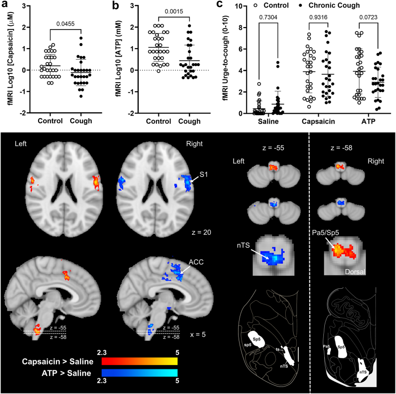
Background: Chronic cough is a prevalent and difficult to treat condition often accompanied by cough hypersensitivity, characterised by cough triggered from exposure to low level sensory stimuli. The mechanisms underlying cough hypersensitivity may involve alterations in airway sensory nerve responsivity to tussive stimuli which would be accompanied by alterations in stimulus-induced brainstem activation, measurable with functional magnetic resonance imaging (fMRI).

Methods: We investigated brainstem responses during inhalation of capsaicin and adenosine triphosphate (ATP) in 29 participants with chronic cough and 29 age- and sex-matched controls. Psychophysical testing was performed to evaluate individual sensitivities to inhaled stimuli and fMRI was used to compare neural activation in participants with cough and control participants while inhaling stimulus concentrations that evoked equivalent levels of urge-to-cough sensation.

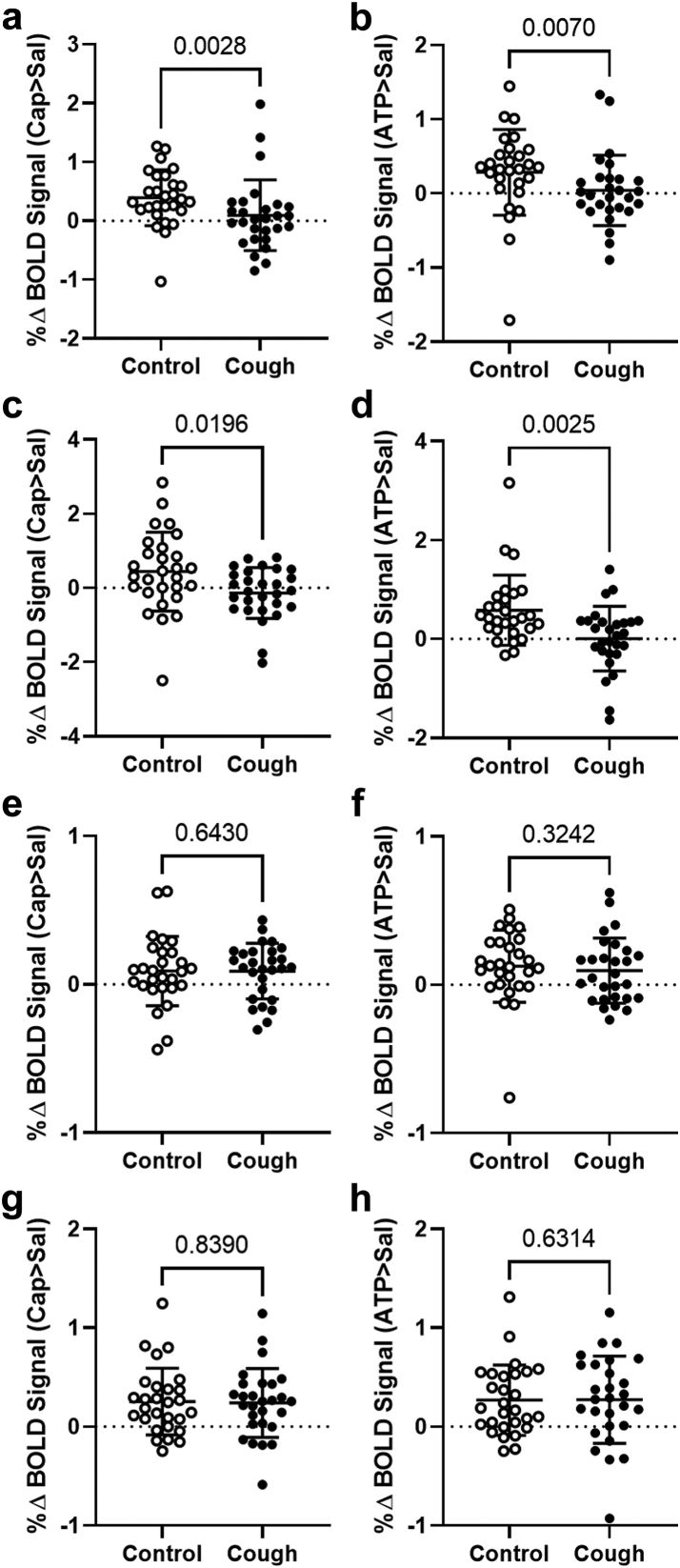
Findings: Participants with chronic cough were significantly more sensitive to inhaled capsaicin and ATP and showed a change in relationship between urge-to-cough perception and cough induction. When urge-to-cough levels were matched, participants with chronic cough displayed significantly less neural activation in medullary regions known to integrate airway sensory inputs. By contrast, neural activations did not differ significantly between the two groups in cortical brain regions known to encode cough sensations whereas activation in a midbrain region of participants with chronic cough was significantly increased compared to controls.

Interpretation: Cough hypersensitivity in some patients may occur in brain circuits above the level of the medulla, perhaps involving midbrain regions that amplify ascending sensory signals or change the efficacy of central inhibitory control systems that ordinarily serve to filter sensory inputs.

Funding: Supported in part by a research grant from Investigator-Initiated Studies Program of Merck Sharp & Dohme Pty Ltd. The opinions expressed in this paper are those of the authors and do not necessarily represent those of Merck Sharp & Dohme (Australia) Pty Ltd.

Keywords: ATP; Brain imaging; Brainstem; Cough; Purinergic; Sensitisation; Vagal sensory.


Conflict of interest statement

Declaration of interests SBM reports receiving grants from the National Health and Medical Research Council (NHMRC) of Australia and the Australian Research Council (ARC), Merck, Bellus Health and Reckitt Benkiser, and remuneration for consultancy from Merck, Trevi Therapeutics, Reckitt Benkiser and Nerre Therapeutics, has served on advisory committees for Reckitt Benkiser and has received payment from Reckitt Benkiser for assistance with manuscript writing. KFC reports research grants from Merck and GSK, remuneration for lectures from Novartis and AstraZeneca; has served on advisory boards for Roche, Merck, Reckitt Benckiser, and Shionogi & Co., Ltd., and a Data Safety Monitoring Board for Nocion. LMG reports research grants from Bayer AG, Bellus Health, Chiesi, Merck, and Shionogi, remuneration for lectures from Bayer AG, Bellus Health, Chiesi, GlaxoSmithKline, Merck, and Shionogi, remuneration for consultancy from Bayer AG, Bellus Health, Chiesi, Merck, NeRRe Therapeutics, Nocion Therapeutics, and Shionogi, and has served on advisory committees for Applied Clinical Intelligence, Bayer AG, Bellus Health, Chiesi, Merck, NeRRe Therapeutics, Nocion Therapeutics, and Shionogi and on a Data and Safety Monitoring Board for Bayer AG. All other authors declare no relevant conflict of interest.

Figures

Fig. 1
Fig. 1
Summary of screening and recruitment for participants with chroniccough.
Fig. 2
Fig. 2
Overview of fMRI experimental design. (a) Control participants and participants with chronic cough underwent inhaled cough challenge testing during brainstem optimised functional imaging (restricted field of view highlighted by yellow shaping). (b) ATP and capsaicin challenge concentrations were individually tailored to produce behaviourally equivalent levels of urge-to-cough in all participants. Stimuli were delivered across four brain imaging runs, comprising two 24 s blocks each of saline, capsaicin, and ATP inhalation (6 blocks/run in total).
Fig. 3
Fig. 3
Comparison of capsaicin and ATP psychophysical cough indices in control participants and participants with chronic cough. Upper panels show (a) Capsaicin and (b) ATP behavioural thresholds. Lower panels show urge-to-cough ratings provided by control participants and participants with chronic cough while inhaling Cu and C2 concentrations of (c, d) capsaicin and (e, f) ATP. C2-Cu urge-to-cough rating difference scores were calculated for both capsaicin (d) and ATP (f). Cu, Urge-to-cough threshold; C2, stimulus concentration producing two spontaneous coughs; Smax, maximum stimulus concentration tolerated without coughing during 24 s of repeated inhalation. Data points represent individual participants (horizontal line denotes mean and error bars are standard deviation) of n = 29 per group. P values calculated using two-way ANOVA with Šídák's multiple comparison tests (a, b) and unpaired t-tests (c–f).
Fig. 4
Fig. 4
Capsaicin and ATP inhalation during fMRI scanning. (a) Capsaicin and (a) ATP stimulus concentrations used for control participants and participants with chronic cough during fMRI scanning to achieve (c) equivalent urge-to-cough experiences for control and chronic cough groups for each test stimulus. Data points represent individual participants (horizontal line denotes mean and error bars are standard deviation) of n = 28 per group. P values calculated using unpaired t-tests (a, b) and two-way ANOVA with Šídák's multiple comparison tests. Images show cluster-corrected Blood Oxygen Level Dependent (BOLD) signal responses in the cerebral hemispheres and brainstem reflecting the Capsaicin > Saline (red) and ATP > Saline (blue) activation patterns common for all participants in the study (n = 56). Slices and region co-ordinates displayed were chosen based on the outcomes of our prior fMRI studies.,,, Atlas drawings show the expected location of brainstem regions of interest (modified from35). ACC, anterior cingulate cortex; nTS, nucleus of the solitary tract; Pa5, paratrigeminal nucleus; Sp5, spinal trigeminal nucleus; sp5, spinal trigeminal tract; S1, primary somatosensory cortex; ts, solitary tract.
Fig. 5
Fig. 5
Medullary and cortical BOLD signal changes during inhalation of tussive substances in control participants and participants with chronic cough. Percentage Blood Oxygen Level Dependent (BOLD) signal changes for Capsaicin > Saline (left column graphs, a, c, e, g) and ATP > Saline (right column graphs, b, d, f, h) extracted from regions of interest in the brainstem and cortex of control participants and participants with chronic cough. (a, b) Nucleus of the solitary tract; (c, d) Paratrigeminal nucleus; (e, f) Anterior cingulate cortex; (g, h) Primary sensory cortex. Data points represent individual participants (horizontal line denotes mean and error bars are standard deviation) of n = 28 per group. P values calculated using Mann–Whitney U tests. Abbreviations: ATP, adenosine triphosphate; CAP, capsaicin; SAL, saline.
Fig. 6
Fig. 6
Midbrain BOLD signal changes during inhalation of tussive substances in control participants and participants with chronic cough. (a) Activation map (red) for the contrast Cough Participant > Control Participant, Capsaicin > Saline. Percentage Blood Oxygen Level Dependent (BOLD) signal changes extracted from the activated region (arrow) show that participants with chronic cough have significantly higher signal levels compared to controls for (b) Capsaicin > Saline and (c) ATP > Saline contrasts. Data points represent individual participants (horizontal line denotes mean and error bars are standard deviation) of n = 28 per group. P values calculated using Mann–Whitney U tests. Abbreviations: ATP, adenosine triphosphate; CAP, capsaicin; SAL, saline.

References

    1. Song W.J., Chang Y.S., Faruqi S., et al. The global epidemiology of chronic cough in adults: a systematic review and meta-analysis. Eur Respir J. 2015;45(5):1479–1481. - PubMed
    1. Chung K.F., McGarvey L., Song W.J., et al. Cough hypersensitivity and chronic cough. Nat Rev Dis Primers. 2022;8(1):45. - PMC - PubMed
    1. Morice A.H., Faruqi S., Wright C.E., Thompson R., Bland J.M. Cough hypersensitivity syndrome: a distinct clinical entity. Lung. 2011;189(1):73–79. - PubMed
    1. Kum E., Guyatt G.H., Devji T., et al. Cough symptom severity in patients with refractory or unexplained chronic cough: a systematic survey and conceptual framework. Eur Respir Rev. 2021;30(161) - PMC - PubMed
    1. Kum E., Guyatt G.H., Munoz C., et al. Assessing cough symptom severity in refractory or unexplained chronic cough: findings from patient focus groups and an international expert panel. ERJ Open Res. 2022;8(1) doi: 10.1183/23120541.00667-2021. - DOI - PMC - PubMed