Clinical Trial
. 2024 Jan 20;15(1):646.
doi: 10.1038/s41467-024-44776-4.

Engineering tumor-colonizing E. coli Nissle 1917 for detection and treatment of colorectal neoplasia

Affiliations
Clinical Trial

Engineering tumor-colonizing E. coli Nissle 1917 for detection and treatment of colorectal neoplasia

Candice R Gurbatri et al. Nat Commun. .

Abstract

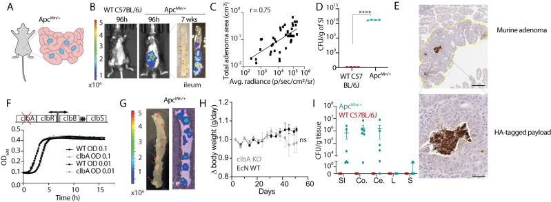
Bioengineered probiotics enable new opportunities to improve colorectal cancer (CRC) screening, prevention and treatment. Here, first, we demonstrate selective colonization of colorectal adenomas after oral delivery of probiotic E. coli Nissle 1917 (EcN) to a genetically-engineered murine model of CRC predisposition and orthotopic models of CRC. We next undertake an interventional, double-blind, dual-centre, prospective clinical trial, in which CRC patients take either placebo or EcN for two weeks prior to resection of neoplastic and adjacent normal colorectal tissue (ACTRN12619000210178). We detect enrichment of EcN in tumor samples over normal tissue from probiotic-treated patients (primary outcome of the trial). Next, we develop early CRC intervention strategies. To detect lesions, we engineer EcN to produce a small molecule, salicylate. Oral delivery of this strain results in increased levels of salicylate in the urine of adenoma-bearing mice, in comparison to healthy controls. To assess therapeutic potential, we engineer EcN to locally release a cytokine, GM-CSF, and blocking nanobodies against PD-L1 and CTLA-4 at the neoplastic site, and demonstrate that oral delivery of this strain reduces adenoma burden by ~50%. Together, these results support the use of EcN as an orally-deliverable platform to detect disease and treat CRC through the production of screening and therapeutic molecules.


Conflict of interest statement

T.D., N.A., D.L.W., S.L.W. and C.R.G. have financial interest in GenCirq Inc. T.D., D.L.W., S.L.W. and C.R.G. have filed a provisional patent application (“Colorectal Cancer Screening, Prevention, And Treatment With Engineered Probiotics”) with the US Patent and Trademark Office related to this manuscript. The remaining authors have no other competing interests.

Figures

Fig. 1
Fig. 1. Adenoma colonization of E. coli Nissle 1917 (EcN) in a genetically-engineered mouse model of CRC.
A Schematic of spontaneous intestinal adenomas in ApcMin/+ model. 12-week-old ApcMin/+ mice were gavaged twice, 3–4 days apart with EcN expressing luxCDABE luciferase cassette (EcN-lux). B EcN-lux was visualized using an IVIS for bioluminescence in vivo 96 h post dosing. After 7 weeks, mice were sacrificed, intestinal tissue was excised and imaged ex vivo for bioluminescence. Red arrows point to macroadenomas on distal intestinal tissue (n = 20 WT, n = 25 ApcMin/+). C Plot where x-axis is the bioluminescence signal measured from ex vivo images of dissected intestinal tissue and y-axis is the total adenoma area in matched tissue sections as measured in H&E-stained images (n = 35 intestinal sections, r = 0.75, Spearman correlation coefficient). D, E In separate cohorts, mice were dosed with EcN-lux or EcN producing an HA-tagged protein and D sacrificed at 1 week post dosing. Intestinal tissue was homogenized and plated on antibiotic-selective plates for EcN-lux to quantify colony-forming-units (CFU) per gram of tissue (n = 4 WT, n = 4 ApcMin/+, ****p < 0.0001) or E sacrificed at 4 weeks post dosing and intestinal tissue was paraffin-embedded, sectioned and stained by anti-HA immunohistochemistry. Dark brown stain depicts HA-tagged protein produced by EcN in adenomas. Scale bars represent 200 μm (top) and 50 μm (bottom). F Schematic (Top) of colibactin-encoding operon in EcN whereby clbA is knocked out and colibactin production is disrupted. Plate reader experiment of strain variant growth kinetics at 0.1 and 0.01 seeding OD. GI 12-week-old ApcMin/+ mice and WT littermates were gavaged twice, 3–4 days apart with 109 CFU bioluminescent EcN-lux or EcNΔclbA-lux. G After 1 week, mice were sacrificed, intestinal tissue was excised and ex vivo imaged for bioluminescence. Red arrows point to macroadenomas on distal small intestinal tissue (representative image from n = 5 mice). H Body weight of both EcN-lux and EcNΔclbA-lux-treated mice were tracked over the course of the experiment (n = 3 EcN-lux, n = 4 EcNΔclbA-lux, two-way ANOVA, ns, not significant). I In a separate cohort one week post dosing, mice were sacrificed, small intestine (SI), colon (Co), cecum (Ce), liver (L), and spleen (S) were harvested, homogenized, and plated on antibiotic selective plates to recover EcNΔclbA-lux per gram of tissue (n = 3 WT, n = 6 ApcMin/+ mice per group). All error bars represent S.E.M. Source data are provided as a Source data file.
Fig. 2
Fig. 2. Tumor colonization of E. coli Nissle 1917 (EcN) in orthotopic mouse models and human CRC patients.
A Schematic of experimental timeline for MSS CRC model. Tumor growth was monitored by colonoscopy, with animals orally dosed twice with EcN-lux or PBS. B Imaging 5 days after last dose for bioluminescence, with C luminescence quantified in organs ex vivo (L, liver, S, spleen, and NC, normal colon n = 17, PT, proximal tumor, DT distal tumor, n = 12, PBS tumors n = 6). D Tissues were homogenized, plated on antibiotic-selective plates, and quantified for CFU per gram (n = 17 from EcN-dosed non-tumor mice and EcN-dosed tumor mice n = 6, PBS-dosed tumor mice n = 4). E, F Representative images of orthotopic CRC from mice dosed as (A), with serial sections showing (E) EcN-lux specific location by RNAscope in situ hybridization for lux (brown, scale bar 20 μm) and (F) immunofluorescent staining for Hypoxyprobe (red) and lipopolysaccharide (LPS, green, scale bar 10 μm, n = 5 mice). G Schematic of experimental timeline for MSI CRC model. Tumor and non-tumor bearing control animals orally dosed thrice with EcN-lux. H Imaging 5 days after last dose with luminescence quantified in organs ex vivo and I tissues homogenized, plated on antibiotic-selective plates, and quantified for CFU per gram (n = 4 for both tumor bearing and no-tumor control groups). J Schematic of human clinical trial. K Matched normal and tumor tissue homogenates from CRC patients administered placebo (n = 2) or Mutaflor (trade name for EcN) (n = 6) for 2 weeks. Tissue samples were used to inoculate liquid culture for microbial enrichment. DNA isolated from culture was subjected to qPCR with an EcN specific assay. Mean value of 4 technical replicates shown per sample, box in the plot minima–maxima is 25th–75th percentile, whiskers at lowest and highest values, bar at median per group. Red dashed line depicts the lower limit of detection of each assay based on standard curve dilution series, dot points above the line have detectable PCR amplicon signal. No EcN signal was detected in E. coli control (ATCC 9522), no template (NTC), or buffer only DNA prep controls. C, D, I ****p < 0.0001, one-way ANOVA with Tukey’s multiple comparisons. H ****p < 0.0001, two-way ANOVA, Fischer’s LSD test). K *p = 0.03, two-way paired t-test. Figure 2J was created with BioRender.com. Source data are provided as a Source data file.
Fig. 3
Fig. 3. Engineering of stool and urine-based EcN platform for non-invasive adenoma tracking.
A Schematic of orally-delivered EcN probiotic to be detected in fecal matter by quantifying colony-forming units or urine by metabolite quantification. B 15-week-old ApcMin/+ mice were dosed orally 3–4 days apart with EcN and one stool pellet was collected at 3, 7, 24, 48, and 72 h after last dose, homogenized, plated on antibiotic-selective plates and quantified for CFU (n = 5 mice per group). C Schematic of the genetic construct where the isochoristmate synthase (ICS) and pchB genes result in salicylate production (Top). These enzymes were cloned onto plasmids and transformed into EcN or a metabolically engineered EcN strain (EcNATT). D, E Overnight cultures of these strain variants were optical density-matched and LC-MS was used to probe for salicylate presence between D plasmid-encoded EcN ATT variants (n = 6 biological replicates per group) and E selected variants between the EcN and EcNATT strains (n = 3 biological replicates per group, two-way ANOVA, Holm-Sidak post test ***p = 0.0001, ****p < 0.0001). All samples were normalized to an internal isotope-labeled D4-salicylate standard. F, G 15-week-old ApcMin/+ mice were dosed with 109 EcNATT-EntC-ColE1, F stool was collected at 48 h, homogenized, and plated on selective plates to quantify recoverable bacteria (n = 3 mice per group, Er) and recoverable bacteria retaining the salicylate-encoded plasmid (Kan) and G urine was collected 24 h and 48 h after dosing and salicylate quantified using LC-MS in WT and ApcMin/+ mice (n = 3 mice per group, ns = not significant, *p = 0.0228 two-way ANOVA with Holm-Sidak post test). All error bars represent S.E.M. Source data are provided as a Source data file.
Fig. 4
Fig. 4. Treatment with EcN engineered to produce immunotherapeutics reduces adenoma burden and modifies the tumor-immune microenvironment.
A Schematic of orally-delivered EcN probiotic engineered to lyse and produce immunotherapeutic proteins in situ (top) and schematic of dosing regimen (bottom). B, C 15-week-old ApcMin/+ mice were dosed twice within 3–4 days and then with PBS (Unt), EcN genomically encoding a lysis circuit (SLIC) or SLIC producing granulocyte-macrophage colony-stimulating factor (GM-CSF), and blocking nanobodies against PD-L1 and CTLA-4 targets (SLIC-3). One month after dosing, mice were sacrificed, intestines were bisected, swiss-rolled, paraffin-embedded, sectioned, stained with hemotoxin and eosin and quantified for B overall tumor area (black: female mice; gray: male mice), n = 5 mice (Untreated), n = 8 (SLIC), n = 10 (SLIC-3) and C tumor area along the intestine, D, duodenum, PJ, proximal jejunum, DJ, distal jejunum, I, ileum (*p = 0.0215, **p = 0.0008, ***p = 0.001, ns, not significant, ordinary one-way ANOVA test with Holm-sidak multiple comparisons test, n = 5 mice (Untreated), n = 7 (SLIC), n = 7 (SLIC-3). D Representative H&E-stained histology images of SLIC and SLIC-3 treated mice. EG Using immunohistochemical (IHC) techniques, intestinal tissue sections from each mouse groups were stained and quantified for E CD3+, n = 3 mice, 113 polyps (SLIC-3), n = 4 mice, 107 polyps (SLIC), n = 3 mice, 131 polyps (Untreated), ****p < 0.0001 ordinary one-way ANOVA with Tukey post-test), F CD8+, n = 3 mice, 111 polyps (SLIC-3), n = 4 mice, 141 polyps (SLIC), n = 3 mice, 112 polyps (Untreated), ****p < 0.0001 ordinary one-way ANOVA with Tukey post-test), and G granzymeB+ cells, n = 3 mice, 124 polyps (SLIC-3), n = 3 mice, 120 polyps (SLIC), n = 3 mice, 112 polyps (Untreated), ****p < 0.0001 ordinary one-way ANOVA with Tukey post-test). Representative IHC images of all three markers are shown beside their respective plots with positive staining depicted as brown puncta in SLIC and SLIC-3-treated mice. Scale bars represent 200 μm. All error bars represent S.E.M. Source data are provided as a Source data file.

References

    1. Mazzolini, R. et al. Engineered live bacteria suppress Pseudomonas aeruginosa infection in mouse lung and dissolve endotracheal-tube biofilms. Nat. Biotechnol.41, 1089–1098 (2023). - PMC - PubMed
    1. Russell BJ, et al. Intestinal transgene delivery with native E. coli chassis allows persistent physiological changes. Cell. 2022;185:3263–3277. e3215. doi: 10.1016/j.cell.2022.06.050. - DOI - PMC - PubMed
    1. Whitfill T, Oh J. Recoding the metagenome: microbiome engineering in situ. Curr. Opin. Microbiol. 2019;50:28–34. doi: 10.1016/j.mib.2019.09.005. - DOI - PubMed
    1. Forbes NS. Engineering the perfect (bacterial) cancer therapy. Nat. Rev. Cancer. 2010;10:785–794. doi: 10.1038/nrc2934. - DOI - PMC - PubMed
    1. Chien T, Doshi A, Danino T. Advances in bacterial cancer therapies using synthetic biology. Curr. Opin. Syst. Biol. 2017;5:1–8. doi: 10.1016/j.coisb.2017.05.009. - DOI - PMC - PubMed