. 2024 Jan 20;15(1):76.
doi: 10.1038/s41419-024-06465-4.

The LKB1-TSSK1B axis controls YAP phosphorylation to regulate the Hippo-YAP pathway

Affiliations

The LKB1-TSSK1B axis controls YAP phosphorylation to regulate the Hippo-YAP pathway

Cho-Long Kim et al. Cell Death Dis. .

Erratum in

Abstract

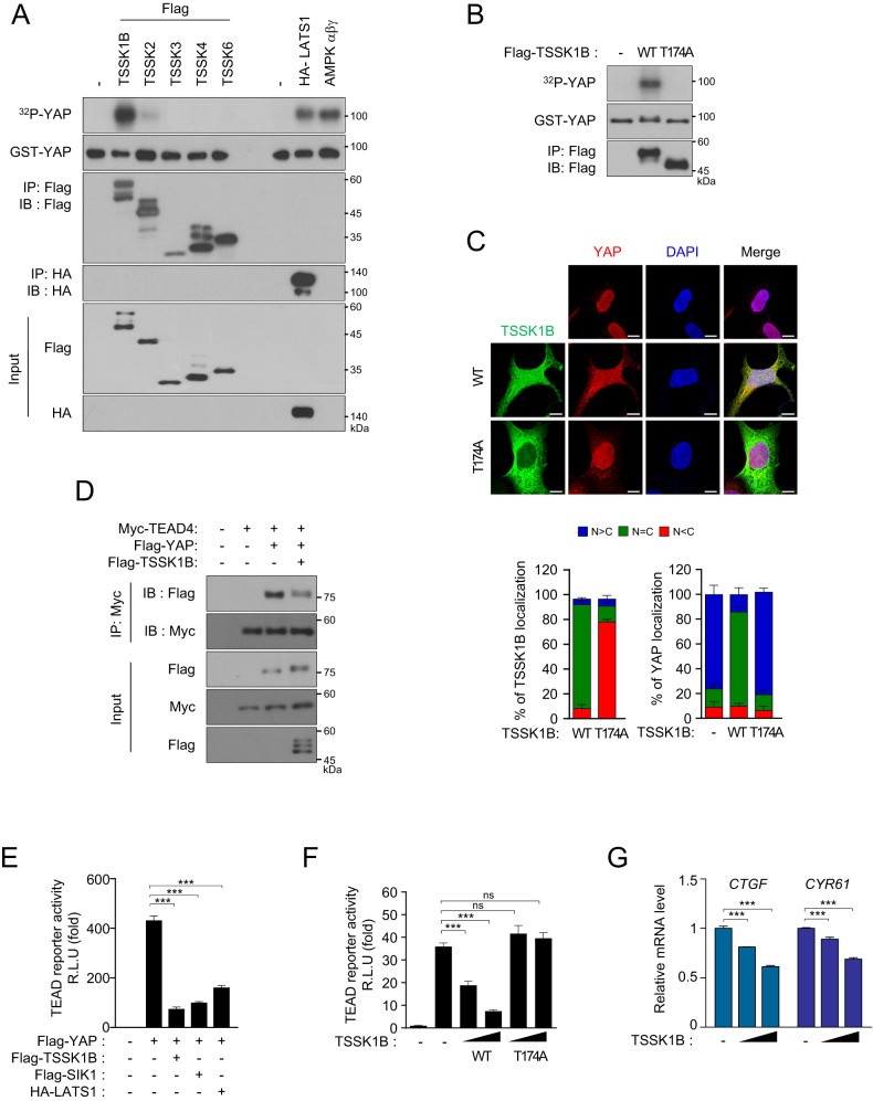
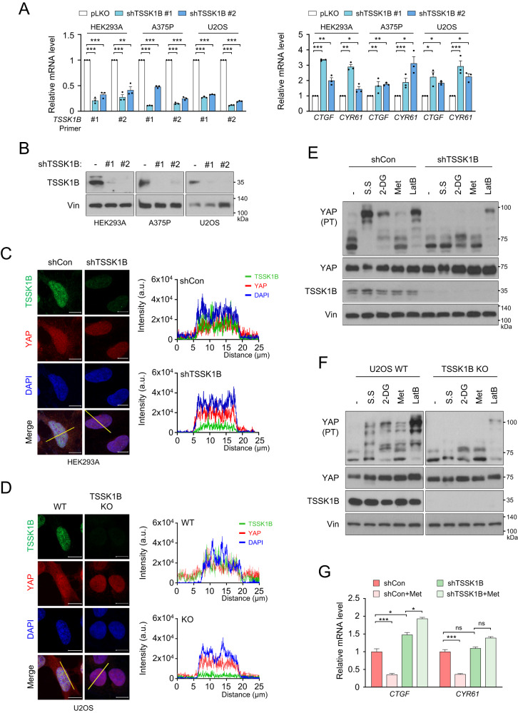
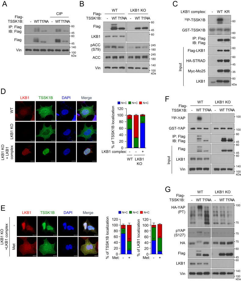
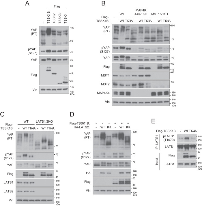
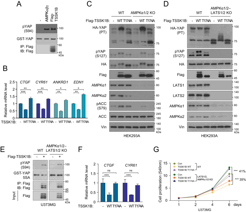
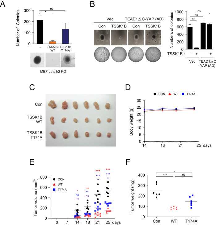
The Hippo pathway's main effector, Yes-associated protein (YAP), plays a crucial role in tumorigenesis as a transcriptional coactivator. YAP's phosphorylation by core upstream components of the Hippo pathway, such as mammalian Ste20 kinase 1/2 (MST1/2), mitogen-activated protein kinase kinase kinase kinases (MAP4Ks), and their substrate, large tumor suppressor 1/2 (LATS1/2), influences YAP's subcellular localization, stability, and transcriptional activity. However, recent research suggests the existence of alternative pathways that phosphorylate YAP, independent of these core upstream Hippo pathway components, raising questions about additional means to inactivate YAP. In this study, we present evidence demonstrating that TSSK1B, a calcium/calmodulin-dependent protein kinase (CAMK) superfamily member, is a negative regulator of YAP, suppressing cellular proliferation and oncogenic transformation. Mechanistically, TSSK1B inhibits YAP through two distinct pathways. Firstly, the LKB1-TSSK1B axis directly phosphorylates YAP at Ser94, inhibiting the YAP-TEAD complex's formation and suppressing its target genes' expression. Secondly, the TSSK1B-LATS1/2 axis inhibits YAP via phosphorylation at Ser127. Our findings reveal the involvement of TSSK1B-mediated molecular mechanisms in the Hippo-YAP pathway, emphasizing the importance of multilevel regulation in critical cellular decision-making processes.


Conflict of interest statement

The authors declare no competing interests.

Figures

Fig. 1
Fig. 1. TSSK1B inhibits the transcriptional activity of YAP through phosphorylation.
A Flag-TSSK kinase members and HA-LATS1 were expressed in HEK293A cells, and immunoprecipitated kinases and purified AMPKαβγ were used for the kinase reaction with the substrate GST-YAP prepared from Escherichia coli. Phospho-YAP (pYAP) was analyzed using 32P autoradiography, and GST-YAP was confirmed by immunoblotting. - : empty vector for control. B Flag-TSSK1Β wild-type (WT) or mutant (T174A) construct was immunoprecipitated using anti-Flag Ab. Immunoprecipitated kinases were used for the kinase reaction with GST-YAP. pYAP was analyzed using 32P autoradiography. C HEK293A cells were transiently transfected with Flag-TSSK1B WT or mutant (T174A) construct and were subjected to immunofluorescence staining for TSSK1Β (green) and YAP (red); 4’,6-diamidino-2-phenylindole (DAPI) (blue) was used for nuclear staining. The nuclear (N)–cytoplasmic (C) ratio of YAP was analyzed in three randomly selected fields from three independent experiments (n = 100 cells per field). Scale bars, 10 µm. D Flag-TSSK1B along with Flag-YAP and Myc-TEAD4 were expressed in HEK293A cells, and Myc-TEAD4 was immunoprecipitated using anti-Myc Ab followed by immunoblotting. E Constructs of different kinases were co-transfected with YAP, GAL4-TEAD4, 5xUAS luciferase reporter, or Renilla into HEK293A cells. After 48 h, the luminescent signal controlled by YAP–TEAD was measured and normalized to Renilla luciferase activity. Error bars indicate the standard error of the mean (SEM) from three separate points in one experiment (n = 3). ***p < 0.001. Student’s t-test was used for statistical analysis. F Flag-TSSK1B WT or mutant (T174A) construct was co-transfected with YAP, GAL4-TEAD4, 5xUAS luciferase reporter, or Renilla into HEK293A cells. After 48 h, the luminescent signal controlled by YAP–TEAD was measured and normalized to Renilla luciferase activity. Error bars indicate mean ± SEM (n = 3). ***p < 0.001. NS: Not significant. Student’s t-test was used for statistical analysis. G HEK293A cells were transiently transfected with the indicated plasmid. The mRNA levels of CTGF and CYR61 were determined using quantitative real-time polymerase chain reaction (qRT-PCR) and normalized to hypoxanthine-guanine phosphoribosyltransferase 1 (HPRT1) mRNA levels. Error bars indicate mean ± SEM (n = 3). ***p < 0.001. Student’s t-test was used for statistical analysis.
Fig. 2
Fig. 2. Deletion of TSSK1B reduces YAP phosphorylation and elevates YAP activity.
A HEK293A, A375P, and U2OS cells were treated with shCon and shTSSK1B and qRT-PCR was performed using specific primer pairs to quantify the mRNA levels of TSSK1B, CTGF, and CYR61. The measurements were normalized with respect to the HPRT1 mRNA levels. Error bars depict mean ± SEM (n = 3). ***p < 0.001. **p < 0.01. *p < 0.05. Student’s t-test was used for statistical analysis. B HEK293A, A375P, and U2OS cells were transduced using shCon or shTSSK1B, and knockdown efficiency for TSSK1B was confirmed by immunoblotting. Vin: Vinculin. C HEK293A cells treated with shCon or shTSSK1B were subjected to immunofluorescence staining for TSSK1Β (green) and YAP (red); 4’,6-diamidino-2-phenylindole (DAPI) (blue) was used for nuclear staining. Scale bars, 10 µm. The fluorescence intensity of TSSK1B, YAP, and DAPI was quantified by drawing a line (yellow) of 25 μm in a merged figure using the ZEN 3.5 blue edition program. D Knockout of TSSK1B using CRISPR/Cas9 in U2OS cells was confirmed by immunocytochemistry. Scale bars, 10 µm. The fluorescence intensity of TSSK1B, YAP, and DAPI was quantified by drawing a line (yellow) of 25 μm in a merged figure using ZEN 3.5 blue edition program. E HEK293A cells treated with shCon and shTSSK1B were incubated with serum-free medium (S.S) for 2 h, 25 mM 2-Deoxy glucose (2-DG) for 2 h, 1 mM metformin (Met) for 4 h, or 0.25 μg·ml−1 latrunculin B (LatB) for 30 min, and whole cell lysates were immunoblotted. PT: phos-tag gel. F U2OS WT and TSSK1B KO cells were incubated with serum-free medium (S.S) for 2 h, 25 mM 2-Deoxy glucose (2-DG) for 2 h, 1 mM metformin (Met) for 4 h, or 0.25 μg·ml−1 latrunculin B (LatB) for 30 min. Whole-cell lysates were immunoblotted. PT: phos-tag gel. G shCon- or shTSSK1B-treated HEK293A cells were incubated with 1 mM metformin for 4 h. The mRNA levels of CTGF and CYR61 were determined. The relative unit values for mRNA were normalized to one with respect to that of the control group without metformin treatment. Error bars depict mean ± SEM (n = 3). ***p < 0.001. *p < 0.05. NS: Not significant. Student’s t-test was used for statistical analysis.
Fig. 3
Fig. 3. Identification of LKB1 as the upstream activator of TSSK1B.
A Immunoprecipitated TSSK1B was treated with calf intestinal alkaline phosphatase (CIP) and detected by immunoblotting. The upper bands disappear after CIP treatment indicating that they represent phosphorylated proteins. Vin: Vinculin. B Cell lysates from HEK293A WT or LKB1 KO cells expressing Flag-TSSK1B WT, or mutant (T174A) were subjected to immunoblotting. ACC: Acetyl CoA carboxylase. C Flag-LKB1 WT or KR, HA-STRAD, and Myc-Mo25 were expressed in HEK293A cells and immunoprecipitated LKB1 kinases were used for the kinase reaction with GST-TSSK1B. pTSSK1B was analyzed using 32P autoradiography, and GST-TSSK1B was detected by immunoblotting. D Flag-TSSK1B or Flag-LKB1 along with HA-STRAD and Myc-Mo25 were expressed in HEK293A WT and LKB1 KO cells, whereafter they were subjected to immunofluorescence assay. The nuclear (N)–cytoplasmic (C) ratio of TSSK1B was analyzed in three randomly selected fields from three independent experiments (n = 100 cells per field). Scale bars, 10 µm. E HEK293A LKB1 KO cells were transiently transfected using Flag-TSSK1B or LKB1 along with HA-STRAD and Myc-Mo25 and treated with 1 mM Metformin for 4 h. Cells were subjected to immunofluorescence assay. The nuclear (N)–cytoplasmic (C) ratios of TSSK1B and LKB1 were analyzed in three randomly selected fields from three independent experiments (n = 30 cells per field). Scale bars, 10 µm. F Flag-TSSK1B WT or mutant (T174A) was expressed in HEK293A WT and LKB1 KO, and immunoprecipitated TSSK1B kinases were used for the kinase reaction with GST-YAP. pYAP was analyzed using 32P autoradiography, and GST-YAP was detected by immunoblotting. G HEK293A WT and LKB1 KO cells were transfected with Flag-TSSK1B WT or mutant (T174A) along with HA-YAP, and whole cell lysates were immunoblotted. PT: phos-tag gel.
Fig. 4
Fig. 4. TSSK1B regulates YAP phosphorylation through the Hippo pathway.
A Cell lysates from HEK293A cells expressing Flag-TSSKs were subjected to immunoblotting. YAP phosphorylation status was analyzed using a phos-tag gel to assess the phosphorylation-dependent mobility shift. Vin: Vinculin. PT: phos-tag gel. B HEK293A WT, MAP4K4/6/7 KO, and MST1/2 KO cells were transiently transfected with Flag-TSSK1B WT or mutant (T174A) construct and whole cell lysates were immunoblotted. PT: phos-tag gel. C HEK293A WT and LATS1/2 KO cells were transfected with the indicated plasmids and whole cell lysates were immunoblotted. PT: phos-tag gel. D HEK293A cells were transfected with empty vector (-), Flag-TSSK1B, LATS2 WT, or LATS2 KR mutants. Whole cell lysates were immunoblotted using the indicated Abs. PT: phos-tag gel. E Flag-TSSK1B WT or mutant (T174A) construct was expressed in HEK293A cells and immunoprecipitated using anti-LATS1 Ab. LATS1 phosphorylation levels were detected by immunoblotting using anti-pLATS1 Thr1079 Ab.
Fig. 5
Fig. 5. TSSK1B phosphorylates YAP at Ser94, negatively affecting its transcriptional activity and cell proliferation.
A Immunoprecipitated Flag-TSSK1B and purified AMPKαβγ complexes were used for the kinase reaction with GST-YAP. YAP phosphorylation was analyzed by immunoblotting. B HEK293A LATS1/2 KO cells were transiently transfected with the indicated plasmids. qRT-PCR determined the mRNA levels of CTGF, CYR61, ANKRD1, and END1. Error bars represent mean ± SEM (n = 3). ***p < 0.001. **p < 0.01. *p < 0.05. Student’s t-test was used for statistical analysis. C HEK293A WT and AMPKα1/2 KO cells were transiently transfected with an empty vector (-), Flag-TSSK1B WT, or mutant (T174A) construct, and whole cell lysates were immunoblotted. YAP phosphorylation status was analyzed using a phos-tag gel. PT: phos-tag gel. Vin: Vinculin. D HEK293A WT and AMPKα1/2–LATS1/2 KO cell pools were transiently transfected with the indicated plasmids, and whole cell lysates were immunoblotted. YAP phosphorylation status was analyzed using a phos-tag gel. PT: phos-tag gel. E U373MG WT and AMPKα1/2–LATS1/2 KO cell pools were transfected with Flag-TSSK1B WT and immunoprecipitated using anti-Flag Ab. Immunoprecipitated Flag-TSSK1B was used with GST-YAP 5SA prepared from E. coli. YAP phosphorylation and GST-YAP 5SA were detected by immunoblotting using anti-pYAP Ser94 or anti-GST Abs, respectively. F U373MG WT and AMPKα1/2–LATS1/2 KO cell pools were transiently transfected with the indicated plasmids. qRT-PCR determined the mRNA levels of CTGF and CYR61. Error bars represent mean ± SEM (n = 3). ***p < 0.001. NS: Not significant. Student’s t-test was used for statistical analysis. G pBABE-vector (Con), pBABE-TSSK1Β WT, or mutants (T174A) were stably expressed in U373MG WT and AMPKα1/2–LATS1/2 KO cell pools, and cell proliferation was measured using SRB assay. Values represent mean ± SEM (n = 3). ***p < 0.001. Student’s t-test was used for statistical analysis.
Fig. 6
Fig. 6. TSSK1B suppresses anchorage-independent growth and inhibits tumor growth independent of LATS1/2.
A MEF Lats1/2 KO cells expressing pBABE-vector (-), TSSK1B WT, or mutant (T174A) were assessed for colony formation in soft agar. The representative colony formations were stained with crystal violet. Values represent mean ± SEM (n = 3). *p < 0.05. NS: Not significant. Student’s t-test was used for statistical analysis. Scale bars, 200 µm. B MEF Lats1/2 KO cells expressing pBABE-vector, pBABE-TSSK1B, pPGS-HA-vector, and pPGS-HA-TEAD1ΔC–YAP(AD) were assessed for colony formation in soft agar. MEF Lats1/2 KO expressing TSSK1B exhibited an increase in soft agar colony formation. Error bars depict mean ± SEM (n = 3). **p < 0.01. NS: Not significant. Student’s t-test was used for statistical analysis. Scale bars, 200 µm. CF TSSK1B inhibits tumor growth. C The image of the tumors dissected from BALB/c nude mice with pBABE-vector (Con), TSSK1Β WT, or mutant (T174A) in MEF Lats1/2 KO cells on the 25th day after injection. D In the three groups, the mice’s body weights did not differ significantly. E The tumor growth in the TSSK1B WT xenograft group was significantly lower than that in the control and mutant (T174A) groups. P-value: Con vs. WT (red), Con vs. mutant (T174A) (blue), WT vs. mutant (T174A) (purple). Error bars depict mean ± SEM (n = 6). ***p < 0.001. **p <0.01. *p < 0.05. NS: Not significant. Student’s t-test was used for statistical analysis. F The average tumor weight was lower in the WT group than in the control and mutant (T174A) groups. Error bars depict mean ± SEM (n = 6). ***p < 0.001. *p < 0.05. NS: Not significant. Student’s t-test was used for statistical analysis.

References

    1. Manning SA, Kroeger B, Harvey KF. The regulation of Yorkie, YAP and TAZ: new insights into the Hippo pathway. Development. 2020;147:dev179069. - PubMed
    1. Zheng Y, Pan D. The Hippo Signaling Pathway in Development and Disease. Dev Cell. 2019;50:264–82. - PMC - PubMed
    1. Ma S, Meng Z, Chen R, Guan KL. The Hippo Pathway: Biology and Pathophysiology. Annu Rev Biochem. 2019;88:577–604. - PubMed
    1. Zhao B, Wei X, Li W, Udan RS, Yang Q, Kim J, et al. Inactivation of YAP oncoprotein by the Hippo pathway is involved in cell contact inhibition and tissue growth control. Genes Dev. 2007;21:2747–61. - PMC - PubMed
    1. Liu CY, Zha ZY, Zhou X, Zhang H, Huang W, Zhao D, et al. The hippo tumor pathway promotes TAZ degradation by phosphorylating a phosphodegron and recruiting the SCFbeta-TrCP E3 ligase. J Biol Chem. 2010;285:37159–69. - PMC - PubMed

Publication types

Substances