. 2024 Jan 20;14(1):1817.
doi: 10.1038/s41598-024-52250-w.

Development and validation of cost-effective SYBR Green-based RT-qPCR and its evaluation in a sample pooling strategy for detecting SARS-CoV-2 infection in the Indonesian setting

Affiliations

Development and validation of cost-effective SYBR Green-based RT-qPCR and its evaluation in a sample pooling strategy for detecting SARS-CoV-2 infection in the Indonesian setting

Ratika Rahmasari et al. Sci Rep. .

Abstract

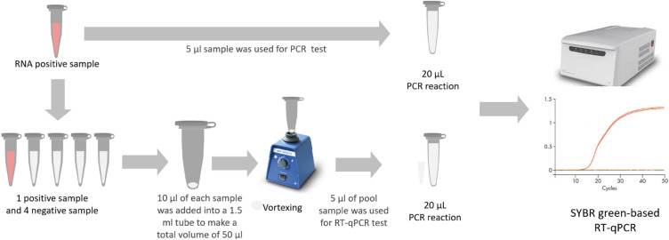
A low-cost SYBR Green-based RT-qPCR method to detect SARS-CoV-2 were developed and validated. Primers targeting a conserved and vital region of the N genes of SARS-CoV-2 were designed. In-silico study was performed to analyse the compatibility of the selected primer pair with Indonesian SARS-CoV-2 genome sequences available from the GISAID database. We determined the linearity of our new assay using serial dilution of SARS-CoV-2 RNA from clinical samples with known virus concentration. The assay was then evaluated using clinically relevant samples in comparison to a commercial TaqMan-based test kit. Finally, we applied the assay in sample pooling strategies for SARS-CoV-2 detection. The SYBR Green-based RT-qPCR method was successfully developed with sufficient sensitivity. There is a very low prevalence of genome variation in the selected N primer binding regions, indicating their high conservation. The validation of the assay using clinical samples demonstrated similar performance to the TaqMan method suggesting the SYBR methods is reliable. The pooling strategy by combining 5 RNA samples for SARS-CoV-2 detection using the SYBR RT-qPCR methods is feasible and provides a high diagnostic yield. However, when dealing with samples having a very low viral load, it may increase the risk of missing positive cases.


Conflict of interest statement

The authors declare no competing interests.

Figures

Figure 1
Figure 1
Location of the N-gene specific primers designed for the study in the SARS-CoV-2 genome.
Figure 2
Figure 2
Schematic illustration of sample pooling technique of SARS-CoV-2 detection.
Figure 3
Figure 3
Characteristics of SYBR Green-based RT-qPCR. (A) Representative amplification curve of the assays with three different primers targeting N gene and GADPH gene as control primer pair, using a positive SARS-CoV-2 RNA sample; (B) melting curve of the products amplified with the N primers and GADPH gene; (C) Distinct PCR products amplified with three different primers and GADPH gene (Line 1, 3, 5, and 7) visualized in 2% agarose. Line 2, 4, 6, and 8 indicate negative control of the assays (Nuclease-free water), respectively (original gel is presented in Supplementary Fig. 2C). Note: the arrows mark the molecular weight of 100 bp and 200 bp (Tiangen, China).
Figure 4
Figure 4
Sequence variants in primer binding regions for N158 primer forward (A) and reverse (B). A total of 998 SARS-CoV-2 genome sequences from infected individuals of Indonesia, including delta (n = 250), omicron and its subvariants BA.1, BA.2, BA.3, BA.4.1, BA.5, BF.5 and XBB.1 (n = 748) submitted from November 2021 until the end of May 2023 were downloaded from the GISAD database (https://www.gisaid.org/). Using Bioedit Sequence Alignment Editor, each primer was aligned to the genome sequences to calculate nucleotide diversity and investigate mismatches to the genome sequence sets. The position and frequency of each variant/mismatch on the primers was then plotted.
Figure 5
Figure 5
Analytical sensitivity of SYBR Green-based RT-qPCR method for SARS-CoV-2 detection. (A) Representative amplification curves for N158 assay targeting N specific gene. Amplification plots (cycle number versus fluorescence) of serially diluted of positive SARS-CoV-2 RNA template (copies/reaction). The RNA concentration of the original sample was measured by the methods described in the previously study (Rahmasari et al., 2022). All seven serially diluted samples, ranging from 107 to 101, were tested in triplicate using the SYBR Green-based RT-qPCR protocol. (B) standard curve of N158 assay. RNA copy number is indicated and plotted against the cycle threshold (Ct) value. The coefficient of determination (R2), Y-intercept, slope, and the efficiency (E) of PCR were calculated. (C) Corresponding melt curve dissociation for amplified product, unique single peaks with Tm around 82.9 °C were generated in all samples. (D) Corresponding RT-qPCR amplicons visualized in 2% agarose gel, amplicon with size of 123 bp were specifically amplified as expected (original gels are presented in Supplementary Fig. 4D).
Figure 6
Figure 6
Validation of the SYBR Green-based RT-qPCR method using a total of 124 clinical samples, comprising 82 positive samples and 42 negative samples. (A) SYBR Green-based RT-qPCR amplification curves for 20 representative positive samples and 10 representative negative samples. (B) Corresponding melt curve dissociation for representative positive and negative samples, unique single peaks with Tm around 82 °C were generated in all positive samples. No nonspecific melt peaks were detected in all negative samples. (C) RT-qPCR amplicon with expected size of 123 bp were specifically amplified for positive samples and no unspecifc amplicon were detected in all negative samples (original gels are presented in Supplementary Fig. 5C).
Figure 7
Figure 7
Comparison of Ct value of pooling strategy. Seventy previously identified positive RNA samples were used, including 36 samples that had tested as weak positives (Ct ≥ 30) and 34 samples that had tested as medium positives (Ct < 30). (A) Ct values distribution of pooled (1/5 samples) and individual samples of medium and weak positive measured with the SYBR Green-based RT-qPCR protocol. (B) Mean Ct value (median, range) of medium positive and (C) weak positive. Original data is presented in Supplementary Fig. 7, respectively.

Similar articles

Cited by

References

    1. Kumar A, Gupta PK, Srivastava A. A review of modern technologies for tackling COVID-19 pandemic. Diabetes Metab. Syndr. 2020;14(4):569–573. doi: 10.1016/j.dsx.2020.05.008. - DOI - PMC - PubMed
    1. Thompson RN. Novel coronavirus outbreak in Wuhan, China, 2020: intense surveillance is vital for preventing sustained transmission in new locations. J. Clin. Med. 2020;9:498. doi: 10.3390/jcm9020498. - DOI - PMC - PubMed
    1. Emrani J, Ahmed M, Jeffers-Francis L, Teleha JC, Mowa N, Newman RH, Thomas MD. SARS-COV-2, infection, transmission, transcription, translation, proteins, and treatment: A review. Int. J. Biol. Macromol. 2021;193(Pt B):1249–1273. doi: 10.1016/j.ijbiomac.2021.10.172. - DOI - PMC - PubMed
    1. Peiris JSM, Lai ST, Poon LLM, Guan Y, Yam LYC, Lim W, et al. Coronavirus as a possible cause of severe acute respiratory syndrome. Lancet. 2003;361:1319–1325. doi: 10.1016/S0140-6736(03)13077-2. - DOI - PMC - PubMed
    1. de Groot RJ, Baker SC, Baric RS, Brown CS, Drosten C, Enjuanes L, et al. Commentary: middle East respiratory syndrome coronavirus (MERS-CoV): announcement of the Coronavirus Study Group. J. Virol. 2013;87:7790–7792. doi: 10.1128/JVI.01244-13. - DOI - PMC - PubMed