

Transcriptomic analysis of glucosidase II beta subunit (GluIIß) knockout A549 cells reveals its roles in regulation of cell adhesion molecules (CAMs) and anti-tumor immunity
- PMID: 38245670
- PMCID: PMC10799456
- DOI: 10.1186/s12864-023-09888-z
Transcriptomic analysis of glucosidase II beta subunit (GluIIß) knockout A549 cells reveals its roles in regulation of cell adhesion molecules (CAMs) and anti-tumor immunity
Abstract
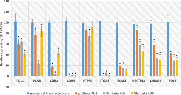
Glucosidase II beta subunit (GluIIß), encoded from PRKCSH, is a subunit of the glucosidase II enzyme responsible for quality control of N-linked glycoprotein folding and suppression of GluIIß led to inhibitory effect of the receptor tyrosine kinase (RTKs) activities known to be critical for survival and development of cancer. In this study, we investigated the effect of GluIIß knockout on the global gene expression of cancer cells and its impact on functions of immune cells. GluIIß knockout lung adenocarcinoma A549 cell line was generated using CRISPR/Cas9-based genome editing system and subjected to transcriptomic analysis. Among 23,502 expressed transcripts, 1068 genes were significantly up-regulated and 807 genes greatly down-regulated. The KEGG enrichment analysis showed significant down-regulation of genes related extracellular matrix (ECM), ECM-receptor interaction, cytokine-cytokine receptor interaction and cell adhesion molecules (CAMs) in GluIIß knockout cells. Of 9 CAMs encoded DEG identified by KEGG enrichment analysis, real time RT-PCR confirmed 8 genes to be significantly down-regulated in all 3 different GluIIß knockout clones, which includes cadherin 4 (CDH4), cadherin 2 (CDH2), versican (VCAN), integrin subunit alpha 4 (ITGA4), endothelial cell-selective adhesion molecule (ESAM), CD274 (program death ligand-1 (PD-L1)), Cell Adhesion Molecule 1 (CADM1), and Nectin Cell Adhesion Molecule 3 (NECTIN3). Whereas PTPRF (Protein Tyrosine Phosphatase Receptor Type F) was significantly decreased only in 1 out of 3 knockout clones. Microscopic analysis revealed distinctively different cell morphology of GluIIβ knockout cells with lesser cytoplasmic and cell surface area compared to parental A549 cells and non-targeted transfected cells.Further investigations revealed that Jurkat E6.1 T cells or human peripheral blood mononuclear cells (PBMCs) co-cultured with GluIIß knockout A549 exhibited significantly increased viability and tumor cell killing activity compared to those co-cultured with non-target transfected cells. Analysis of cytokine released from Jurkat E6.1 T cells co-cultured with GluIIß knockout A549 cells showed significant increased level of angiogenin and significant decreased level of ENA-78. In conclusion, knockout of GluIIß from cancer cells induced altered gene expression profile that improved anti-tumor activities of co-cultured T lymphocytes and PBMCs thus suppression of GluIIß may represent a novel approach of boosting anti-tumor immunity.
Keywords: Cell adhesion molecules (CAMs); Glucosidase II beta subunit; Non-small cell lung cancers (NSCLCs); PRKCSH; T cell; Transcriptomic analysis.
© 2024. The Author(s).
Conflict of interest statement
The authors declare no competing interests.
Figures


Similar articles
-
Navigating PRKCSH's impact on cancer: from N-linked glycosylation to death pathway and anti-tumor immunity.Front Oncol. 2024 Mar 20;14:1378694. doi: 10.3389/fonc.2024.1378694. eCollection 2024. Front Oncol. 2024. PMID: 38571496 Free PMC article. Review.
-
Knockout of glucosidase II beta subunit inhibits growth and metastatic potential of lung cancer cells by inhibiting receptor tyrosine kinase activities.Sci Rep. 2019 Jul 17;9(1):10394. doi: 10.1038/s41598-019-46701-y. Sci Rep. 2019. PMID: 31316108 Free PMC article.
-
Glucosidase II beta subunit (GluIIβ) plays a role in autophagy and apoptosis regulation in lung carcinoma cells in a p53-dependent manner.Cell Oncol (Dordr). 2017 Dec;40(6):579-591. doi: 10.1007/s13402-017-0349-1. Epub 2017 Sep 19. Cell Oncol (Dordr). 2017. PMID: 28929344
-
Integrative RNA profiling of TBEV-infected neurons and astrocytes reveals potential pathogenic effectors.Comput Struct Biotechnol J. 2022 May 30;20:2759-2777. doi: 10.1016/j.csbj.2022.05.052. eCollection 2022. Comput Struct Biotechnol J. 2022. PMID: 35685361 Free PMC article.
-
Leukocyte-cell adhesion: a molecular process fundamental in leukocyte physiology.Immunol Rev. 1990 Apr;114:67-108. doi: 10.1111/j.1600-065x.1990.tb00562.x. Immunol Rev. 1990. PMID: 1973408 Review.
Cited by
-
Navigating PRKCSH's impact on cancer: from N-linked glycosylation to death pathway and anti-tumor immunity.Front Oncol. 2024 Mar 20;14:1378694. doi: 10.3389/fonc.2024.1378694. eCollection 2024. Front Oncol. 2024. PMID: 38571496 Free PMC article. Review.
References
MeSH terms
Substances
Grants and funding
LinkOut - more resources
Full Text Sources
Research Materials
Miscellaneous
