

Epicardial adipose tissue and subclinical incident atrial fibrillation as detected by continuous monitoring: a cardiac magnetic resonance imaging study
- PMID: 38245893
- PMCID: PMC10951027
- DOI: 10.1007/s10554-023-03029-z
Epicardial adipose tissue and subclinical incident atrial fibrillation as detected by continuous monitoring: a cardiac magnetic resonance imaging study
Abstract
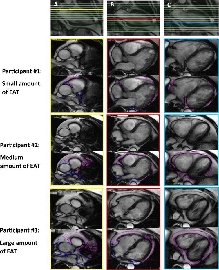
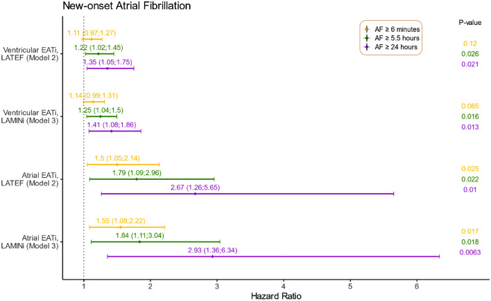
Epicardial adipose tissue (EAT) has endocrine and paracrine functions and has been associated with metabolic and cardiovascular disease. This study aimed to investigate the association between EAT, determined by cardiac magnetic resonance imaging (CMR), and incident atrial fibrillation (AF) following long-term continuous heart rhythm monitoring by implantable loop recorder (ILR). This study is a sub-study of the LOOP study. In total, 203 participants without a history of AF received an ILR and underwent advanced CMR. All participants were at least 70 years of age at inclusion and had at least one of the following conditions: hypertension, diabetes, previous stroke, or heart failure. Volumetric measurements of atrial- and ventricular EAT were derived from CMR and the time to incident AF was subsequently determined. A total of 78 participants (38%) were diagnosed with subclinical AF during a median of 40 (37-42) months of continuous monitoring. In multivariable Cox regression analyses adjusted for age, sex, and various comorbidities, we found EAT indexed to body surface area to be independently associated with the time to AF with hazard ratios (95% confidence intervals) up to 2.93 (1.36-6.34); p = 0.01 when analyzing the risk of new-onset AF episodes lasting ≥ 24 h. Atrial EAT assessed by volumetric measurements on CMR images was significantly associated with the incident AF episodes as detected by ILR.
Keywords: Atrial fibrillation; Cardiac magnetic resonance; Epicardial adipose tissue.
© 2024. The Author(s).
Conflict of interest statement
JHS is a member of Medtronic advisory boards and has received speaker honoraria and research grants from Medtronic in relation to this work and outside the submitted work. SZD is a part-time employee of Vital Beats and has received speaker fees from Pfizer and advisory honoraria from Bristol-Myers Squibb outside the submitted work. AB reports research grants from The Region of Southern Denmark and The Region of Zealand, and Theravance; speaker honoraria from Bayer, Boehringer Ingelheim, and Bristol-Myers Squibb; and a travel grant from Biotronik, outside the submitted work. LK reports speaker honoraria from Novo, AstraZeneca, Novartis, and Boehringer Ingelheim, outside the submitted work. All other authors declare no competing interests.
Figures
Similar articles
-
Left Atrial Late Gadolinium Enhancement is Associated With Incident Atrial Fibrillation as Detected by Continuous Monitoring With Implantable Loop Recorders.JACC Cardiovasc Imaging. 2020 Aug;13(8):1690-1700. doi: 10.1016/j.jcmg.2020.03.024. Epub 2020 Jun 17. JACC Cardiovasc Imaging. 2020. PMID: 32563642
-
The predictive value of epicardial adipose tissue volume assessed by cardiac magnetic resonance for atrial fibrillation in patients with hypertrophic obstructive cardiomyopathy.Int J Cardiovasc Imaging. 2021 Apr;37(4):1383-1393. doi: 10.1007/s10554-020-02092-0. Epub 2021 Jan 3. Int J Cardiovasc Imaging. 2021. PMID: 33392874
-
Association of epicardial adipose tissue and left atrial size on non-contrast CT with atrial fibrillation: the Heinz Nixdorf Recall Study.Eur Heart J Cardiovasc Imaging. 2014 Aug;15(8):863-9. doi: 10.1093/ehjci/jeu006. Epub 2014 Feb 4. Eur Heart J Cardiovasc Imaging. 2014. PMID: 24497517
-
Epicardial adipocytes in the pathogenesis of atrial fibrillation: An update on basic and translational studies.Front Endocrinol (Lausanne). 2023 Mar 20;14:1154824. doi: 10.3389/fendo.2023.1154824. eCollection 2023. Front Endocrinol (Lausanne). 2023. PMID: 37020587 Free PMC article. Review.
-
Epicardial adipose tissue, obesity, and the occurrence of atrial fibrillation: an overview of pathophysiology and treatment methods.Expert Rev Cardiovasc Ther. 2022 Apr;20(4):307-322. doi: 10.1080/14779072.2022.2067144. Epub 2022 Apr 27. Expert Rev Cardiovasc Ther. 2022. PMID: 35443854 Review.
Cited by
-
2025 THRS Expert Consensus of Imaging Assessment in Atrial Fibrillation. Developed by the Task Force of the Taiwan Heart Rhythm Society (THRS) Imaging Committee. Endorsed by the Taiwan Society of Cardiology (TSOC).Acta Cardiol Sin. 2025 Jul;41(4):446-477. doi: 10.6515/ACS.202507_41(4).20250526A. Acta Cardiol Sin. 2025. PMID: 40740204 Free PMC article.
-
Screening for atrial fibrillation: the role of CHA2DS2-VASc and atrial fibrillation burden.Eur Heart J Suppl. 2024 Jul 31;26(Suppl 4):iv41-iv49. doi: 10.1093/eurheartjsupp/suae078. eCollection 2024 Jul. Eur Heart J Suppl. 2024. PMID: 39099574 Free PMC article.
References
-
- Hindricks G, et al. 2020 ESC Guidelines for the diagnosis and management of atrial fibrillation developed in collaboration with the European Association for Cardio-Thoracic Surgery (EACTS): the task force for the diagnosis and management of atrial fibrillation of the Europea. Eur Heart J. 2021;42(5):373–498. doi: 10.1093/EURHEARTJ/EHAA612. - DOI - PubMed
MeSH terms
Grants and funding
LinkOut - more resources
Full Text Sources
Medical
