. 2024 Feb:80:101886.
doi: 10.1016/j.molmet.2024.101886. Epub 2024 Jan 19.

Identification of AgRP cells in the murine hindbrain that drive feeding

Affiliations

Identification of AgRP cells in the murine hindbrain that drive feeding

Tomas P Bachor et al. Mol Metab. 2024 Feb.

Abstract

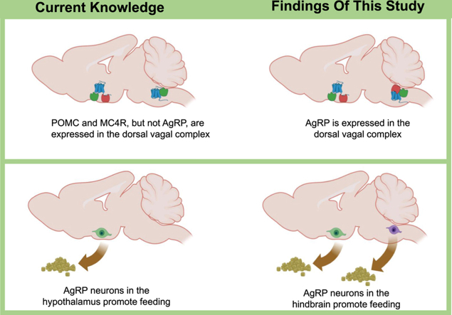
Objective: The central melanocortin system is essential for the regulation of food intake and body weight. Agouti-related protein (AgRP) is the sole orexigenic component of the central melanocortin system and is conserved across mammalian species. AgRP is currently known to be expressed exclusively in the mediobasal hypothalamus, and hypothalamic AgRP-expressing neurons are essential for feeding. Here we characterized a previously unknown population of AgRP cells in the mouse hindbrain.

Methods: Expression of AgRP in the hindbrain was investigated using gene expression analysis, single-cell RNA sequencing, immunofluorescent analysis and multiple transgenic mice with reporter expressions. Activation of AgRP neurons was achieved by Designer Receptors Exclusively Activated by Designer Drugs (DREADD) and by transcranial focal photo-stimulation using a step-function opsin with ultra-high light sensitivity (SOUL).

Results: AgRP expressing cells were present in the area postrema (AP) and the adjacent subpostrema area (SubP) and commissural nucleus of the solitary tract (cNTS) of the mouse hindbrain (termed AgRPHind herein). AgRPHind cells consisted of locally projecting neurons as well as tanycyte-like cells. Food deprivation stimulated hindbrain Agrp expression as well as neuronal activity of subsets of AgRPHind cells. In adult mice that lacked hypothalamic AgRP neurons, chemogenetic activation of AgRP neurons resulted in hyperphagia and weight gain. In addition, transcranial focal photo-stimulation of hindbrain AgRP cells increased food intake in adult mice with or without hypothalamic AgRP neurons.

Conclusions: Our study indicates that the central melanocortin system in the hindbrain possesses an orexigenic component, and that AgRPHind neurons stimulate feeding independently of hypothalamic AgRP neurons.

Keywords: AgRP; Feeding; Hindbrain.


Conflict of interest statement

Declaration of competing interest The authors declare that they have no known competing financial interests or personal relationships that could have appeared to influence the work reported in this paper.

Figures

Image 1
Graphical abstract
Figure 1
Figure 1
AgRP is expressed in the mouse hindbrain and its expression increases with food deprivation. (A) RT-PCR analysis of Agrp mRNA expression in adult mice. PCR products were fractionated on agarose gel. PCR product from cDNA is 378 bp whereas PCR product from genomic DNA is 715 bp. WT: Agrp+/+; Het: Agrp+/−, KO: Agrp−/− mice. (B) qRT-PCR analysis of Agrp mRNA expression in adult mice heterozygous (Het) or null (KO) for AgRP. (C) Picture of the AP marked by Evans blue accumulation and area of microdissection. Cerebellum was removed to expose the AP. (D-E) scRNAseq analyses of the microdissected AP tissues of a P10 mouse, showing cluster plot in D and Agrp-expressing cells in E. (F) Violin plot showing expression of signature genes that mark each cell cluster. (G) scRNAseq analysis of marker genes for neurons, GABAergic and glutamatergic neurons and tanycytes in Agrp cells in microdissected AP-centric tissue from a P10 mouse. (H) snRNAseq analysis of Agrp cells from microdissected AP tissues of chow-fed adult mice [30] (Geo accession number GSE160938). Data were mean of two replicates; each were pools of multiple mice. (I) Agrp mRNA expression in the hindbrain of male and female mice that were ad-lib fed or fasted for 24h. (J) Wide-field images of immunofluorescence analysis with AgRP antibody showing AgRP-positive boutons in mice under fed, 24h fasting or 36h fasting conditions. Scale bar: 100 μm. (K) Confocal Z-scan showing presence of AgRP-positive boutons in the AP of 36h-fasted wild-type (WT) mice but not in AgRP-deficient (KO) mice. Scale bar: 100 μm ∗p < 0.05, ∗∗p < 0.01, ∗∗∗∗p < 0.0001 by students' t-test. AP: area postrema. SubP: subpostrema area. cc: central canal. cNTS: commissural nucleus of solitary tract.
Figure 2
Figure 2
AgRP-expressing cells are present in the AP and cNTS of the mouse hindbrain and their firing rates increase with food deprivation. (A) E15.5 mouse embryos expressing EGFP under the control of AgRP promoter showing expression in the ARC and AP (image ID 78824 is cited with permission from GENSAT). (B) AgRP immunoreactivity in the developing AP at E15.5 in wild-type (WT) and AgRP-deficient (KO) mice. Scale bar: 20 μm. (C) Native fluorescence signals in 300 μm fresh vibratome sections from 10-day-old AgrpCre, RosaTdT/+ mice. Scale bar: 200 μm. (D) Immunofluorescence analysis with DsRed antibody showing tdTomato expression from rostral to caudal AP in adult AgrpCre, RosaTdT/+ mice. Scale bar: 100 μm. (E) Single-scan confocal microscopy of immunoreactivity of neuronal marker HuC/D and tdT in the hindbrain of 9-week-old AgrpCre, RosaTdT/+ mice. Magnified views of the box area in left panel are shown in the 3 panels on the right. Scale bar: 50 μm. (F) Histograms depicting changes in the resting membrane potential (RMP) and action potential frequency (APF) of tdTomato+ neurons in the AP, SubP, and cNTS from ad libitum fed and 24 h fasted AgrpCre, RosaTdT/+ mice. Data are expressed as mean ± SEM. ∗p < 0.05, ∗∗∗p < 0.001, ∗∗∗∗p < 0.0001, determined by unpaired t-test." ARC: arcuate nucleus. DMH: dorsomedial hypothalamus. LHA: lateral hypothalamic area. AP: area postrema. SubP: subpostrema area. cNTS: commissural nucleus of solitary tract. 3V: third ventricle. 4V: fourth ventricle. cc: central canal.
Figure 3
Figure 3
DREADD-mediated activation of AgRP cells in the hindbrain promotes feeding. (A) Mice carrying AgrpCre, RosaLSL-tdT were injected subcutaneously with MSG (3.5 mg/kg) or vehicle at postnatal day 2 (P2) and examined in adulthood. tdTomato+ cells were visualized with DsRed immunoreactivity, followed by single scan confocal microscopy. 3V: third ventricle. ARC: arcuate nucleus. AP: area postrema. Scale bar: 100 μm. (B–C) Mice expressing hM3Dq-DREADD(Gq-DREADD) in AgRP cells (AgrpCre;RosaLSL-hM3Dq, termed AgrphM3Dq) or control mice (AgrpCre or RosaLSL-hM3Dq) were injected with MSG (3.5 mg/kg, s.c.) at P2 to ablate ARC AgRP neurons (ARC-lesioned). n = 6–8 per group. When these mice reached young adulthood (8-week-old), saline was injected on 3 consecutive days, followed by a single CNO (0.5 mg/kg, i.p.) injection. Food intake and body weight were measured at the indicated time periods. (D) ARC-lesioned AgrphM3Dq/+ and control mice were injected with saline and CNO while in the CLAMS chambers. Food intake, RER and energy expenditure were measured. n = 3–4 per group. (E-F) ARC-intact and ARC-lesioned AgrphM3Dq/+ and control mice were treated with CNO, and processed for FOS immunoreactivity 90 min later. Numbers of FOS-positive cells in the ARC and AP were quantified. Scale bar: 100 μm. Data are mean ± SEM. ∗p < 0.05, ∗∗p < 0.01, ∗∗∗p < 0.001, ∗∗∗∗p < 0.0001 by 2-WAY ANOVA with repeated measures and multiple comparisons (panel C and D), 2-WAY ANOVA or student's t-test (panel F).
Figure 4
Figure 4
Transcranial activation of AgRP cells in the hindbrain promotes feeding. (A) Focal transcranial photo-stimulation of AgRPHind cells in mice that expressed the SOUL opsin in AgRP cells (AgrpCre;RosaLSL-SOUL) or in control mice (AgrpCre or RosaLSL-SOUL). Fiber optic cannula was placed above the AP at Bregma −7.5. (B) Food intake was measured at the onset of dark cycle (7 PM) in ad-lib fed ARC-intact male mice (n = 9–10 per group) before and after photo-stimulation (STIM). (C) Food intake was measured at the onset of dark cycle (7 PM) in ad-lib fed 4-month-old ARC-lesioned male mice (n = 4 per group) before and after photo-stimulation. Data are mean ± SEM. ∗∗p < 0.01, by 2-WAY ANOVA with repeated measures and multiple comparisons between control and mutant mice.

Similar articles

Cited by

References

    1. Yeo G.S.H., Chao D.H.M., Siegert A.M., Koerperich Z.M., Ericson M.D., Simonds S.E., et al. The melanocortin pathway and energy homeostasis: from discovery to obesity therapy. Mol Metabol. 2021;48:101206. - PMC - PubMed
    1. Ollmann M.M., Wilson B.D., Yang Y.K., Kerns J.A., Chen Y., Gantz I., et al. Antagonism of central melanocortin receptors in vitro and in vivo by agouti-related protein. Science. 1997;278(5335):135–138. - PubMed
    1. Rossi M., Kim M.S., Morgan D.G., Small C.J., Edwards C.M., Sunter D., et al. A C-terminal fragment of Agouti-related protein increases feeding and antagonizes the effect of alpha-melanocyte stimulating hormone in vivo. Endocrinology. 1998;139(10):4428–4431. - PubMed
    1. Aponte Y., Atasoy D., Sternson S.M. AGRP neurons are sufficient to orchestrate feeding behavior rapidly and without training. Nat Neurosci. 2011;14(3):351–355. - PMC - PubMed
    1. Krashes M.J., Koda S., Ye C., Rogan S.C., Adams A.C., Cusher D.S., et al. Rapid, reversible activation of AgRP neurons drives feeding behavior in mice. J Clin Invest. 2011;121(4):1424–1428. - PMC - PubMed