

Redox State Modulatory Activity and Cytotoxicity of Olea europaea L. (Oleaceae) Leaves Extract Enriched in Polyphenols Using Macroporous Resin
- PMID: 38247497
- PMCID: PMC10812475
- DOI: 10.3390/antiox13010073
Redox State Modulatory Activity and Cytotoxicity of Olea europaea L. (Oleaceae) Leaves Extract Enriched in Polyphenols Using Macroporous Resin
Abstract
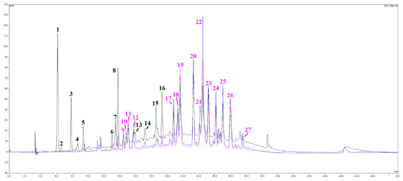
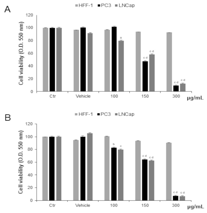
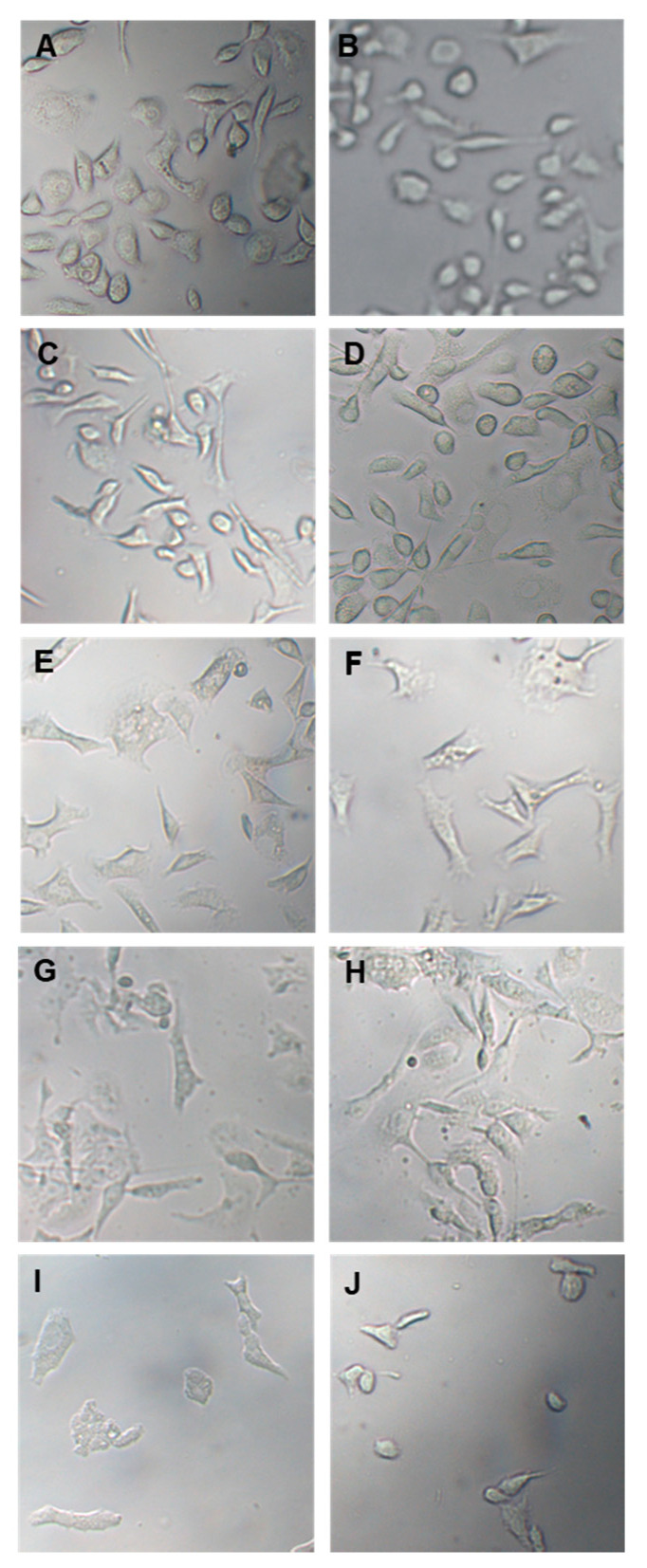
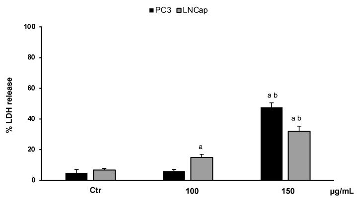
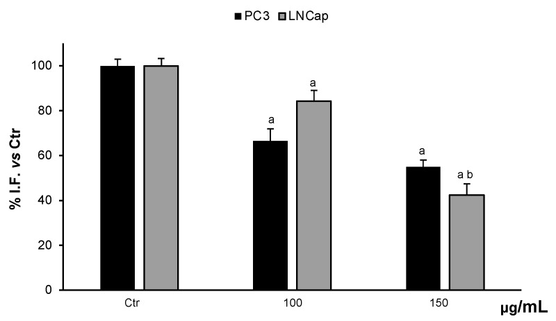
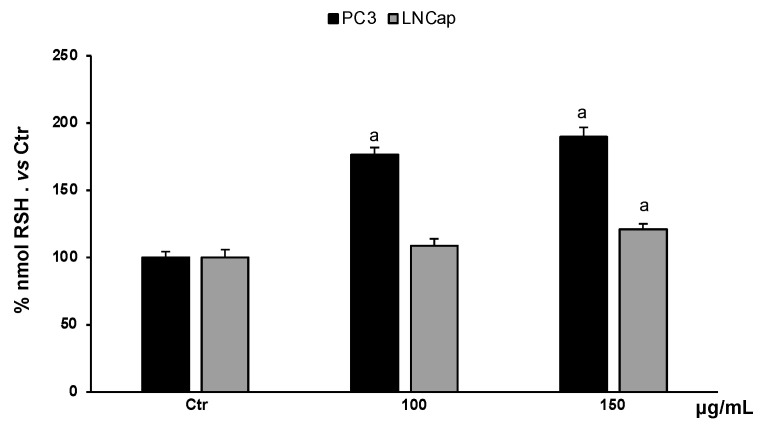
The food products derived from Olea europaea are a fundamental part of the Mediterranean diet, and their health-promoting effects are well known. In this study, we analyzed the phytochemical characteristics, the redox state modulatory activity, and the cytotoxic effect of an olive leaf aqueous extract enriched by macroporous resin on different tumor and normal cell lines (LNCaP, PC3, HFF-1). HPLC-DAD analysis, the Folin-Ciocalteu and aluminum chloride methods confirmed the qualitatively and quantitatively high content of phenolic compounds (130.02 ± 2.3 mg GAE/g extract), and a DPPH assay (IC50 = 100.00 ± 1.8 μg/mL), the related antioxidant activity. The biological investigation showed a significant cytotoxic effect, highlighted by an MTT test and the evident cellular morphological changes, on two prostate cancer cell lines. Remarkably, the extract was practically non-toxic on HFF-1 at the concentrations (100, 150, 300 µg/mL) and exposure times tested. Hence, the results are selective for tumor cells. The underlying cytotoxicity was associated with the decrease in ROS production (55% PC3, 42% LNCaP) and the increase in RSH levels (>50% PC3) and an LDH release assay (50% PC3, 40% LNCaP, established necrosis as the main cell death mechanism.
Keywords: ROS; glutathione; necrosis; oxidative stress; phytochemicals; plant extract; polyphenols; prostate cancer.
Conflict of interest statement
The authors declare no conflict of interest.
Figures
Similar articles
-
Nocellara Del Belice (Olea europaea L. Cultivar): Leaf Extract Concentrated in Phenolic Compounds and Its Anti-Inflammatory and Radical Scavenging Activity.Plants (Basel). 2022 Dec 21;12(1):27. doi: 10.3390/plants12010027. Plants (Basel). 2022. PMID: 36616158 Free PMC article.
-
Waste Autochthonous Tuscan Olive Leaves (Olea europaea var. Olivastra seggianese) as Antioxidant Source for Biomedicine.Int J Mol Sci. 2019 Nov 25;20(23):5918. doi: 10.3390/ijms20235918. Int J Mol Sci. 2019. PMID: 31775339 Free PMC article.
-
Evaluation of the cytotoxicity and anthelmintic activity of Olea europaea (stem and leaves) methanolic extract: in vitro investigation.Helminthologia. 2025 May 24;62(1):8-18. doi: 10.2478/helm-2025-0002. eCollection 2025 Mar. Helminthologia. 2025. PMID: 40443494 Free PMC article.
-
Olive Leaf (Olea europaea L. folium): Potential Effects on Glycemia and Lipidemia.Ann Nutr Metab. 2020;76(1):10-15. doi: 10.1159/000505508. Epub 2020 Jan 3. Ann Nutr Metab. 2020. PMID: 31901903 Review.
-
[Research progress on biological activities of Olea europaea leaf extract].Zhongguo Zhong Yao Za Zhi. 2016 Feb;41(4):613-618. doi: 10.4268/cjcmm20160411. Zhongguo Zhong Yao Za Zhi. 2016. PMID: 28871681 Review. Chinese.
Cited by
-
Protective Effects of Wild Sulla coronaria (Fabaceae) Flowers Phytocomplex in Human Dermal Fibroblasts Stimulated with Interleukin-1β.Plants (Basel). 2024 Sep 30;13(19):2748. doi: 10.3390/plants13192748. Plants (Basel). 2024. PMID: 39409619 Free PMC article.
-
Antidiabetic potential of vanadium complexes combined with olive leaf extracts: a viable approach to reduce metal toxicity.Biometals. 2025 Apr;38(2):683-698. doi: 10.1007/s10534-025-00673-x. Epub 2025 Feb 27. Biometals. 2025. PMID: 40014236 Free PMC article.
-
Origanum majorana Extracts: A Preliminary Comparative Study on Phytochemical Profiles and Bioactive Properties of Valuable Fraction and By-Product.Plants (Basel). 2025 Jul 23;14(15):2264. doi: 10.3390/plants14152264. Plants (Basel). 2025. PMID: 40805613 Free PMC article.
References
-
- ALHaithloul H.A.S., Alotaibi M.F., Bin-Jumah M., Elgebaly H., Mahmoud A.M. Olea europaea leaf extract up-regulates Nrf2/ARE/HO-1 signaling and attenuates cyclophosphamide-induced oxidative stress, inflammation and apoptosis in rat kidney. Biomed. Pharmacother. 2019;111:676–685. doi: 10.1016/j.biopha.2018.12.112. - DOI - PubMed
LinkOut - more resources
Full Text Sources
