Review
. 2024 Jan 18;13(1):120.
doi: 10.3390/antiox13010120.

Diabetic Keratopathy: Redox Signaling Pathways and Therapeutic Prospects

Affiliations
Review

Diabetic Keratopathy: Redox Signaling Pathways and Therapeutic Prospects

Francesco Buonfiglio et al. Antioxidants (Basel). .

Abstract

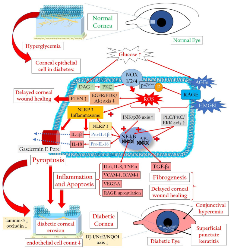
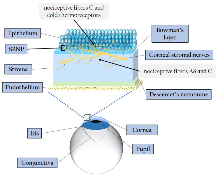
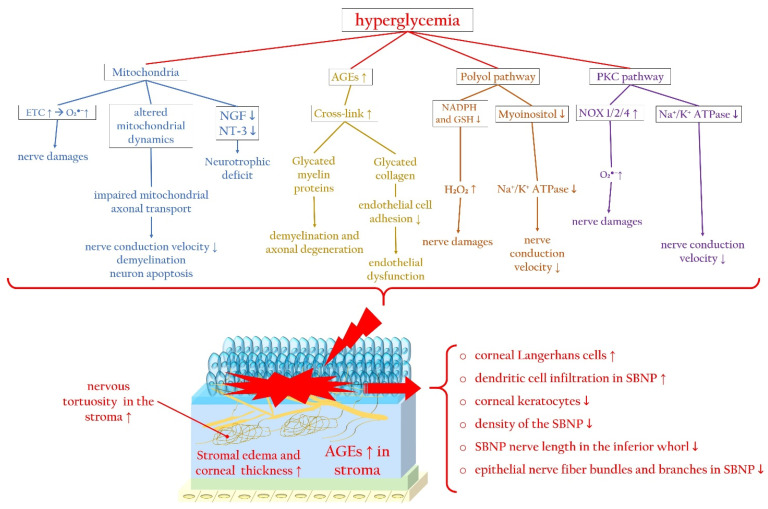
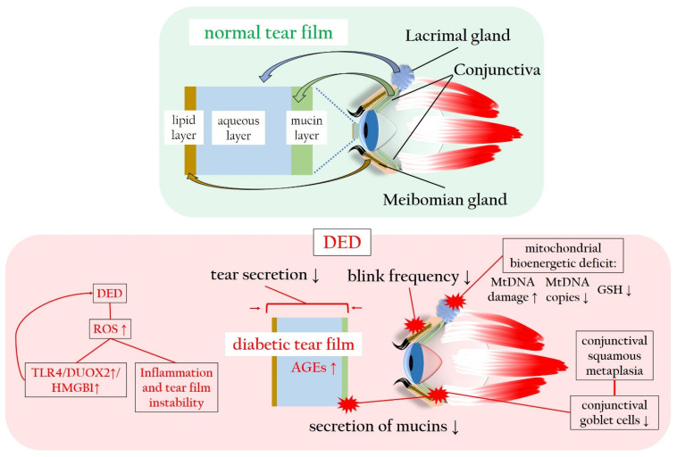
Diabetes mellitus, the most prevalent endocrine disorder, not only impacts the retina but also significantly involves the ocular surface. Diabetes contributes to the development of dry eye disease and induces morphological and functional corneal alterations, particularly affecting nerves and epithelial cells. These changes manifest as epithelial defects, reduced sensitivity, and delayed wound healing, collectively encapsulated in the context of diabetic keratopathy. In advanced stages of this condition, the progression to corneal ulcers and scarring further unfolds, eventually leading to corneal opacities. This critical complication hampers vision and carries the potential for irreversible visual loss. The primary objective of this review article is to offer a comprehensive overview of the pathomechanisms underlying diabetic keratopathy. Emphasis is placed on exploring the redox molecular pathways responsible for the aberrant structural changes observed in the cornea and tear film during diabetes. Additionally, we provide insights into the latest experimental findings concerning potential treatments targeting oxidative stress. This endeavor aims to enhance our understanding of the intricate interplay between diabetes and ocular complications, offering valuable perspectives for future therapeutic interventions.

Keywords: cornea; diabetic complication; keratopathy; molecular; pathways; redox; targets.


Conflict of interest statement

The authors declare no conflicts of interest.

Figures

Figure 1
Figure 1
Slit lamp photograph of a patient with diabetic keratopathy, suffering from dry eye symptoms and recurrent corneal erosions. Fluorescein staining of the ocular surface reveals fine punctate keratitis (yellow dots), which is typical of dry eye disease (DED), highlighted by yellow arrowheads. In the inferior third of the cornea, an epithelial ridge (yellow horizontal line) is visible, indicative of a recently closed erosion, highlighted by yellow arrows.
Figure 2
Figure 2
Cellular signaling mechanisms that lead to the demise of corneal epithelial cells in the context of diabetic keratopathy. AGEs: advanced glycation end products; Akt: Ak strain transforming (also known as protein kinase B); DAG: diacylglycerol; IL: interleukin; DJ-1: parkinsonism-associated deglycase 1; EGFR: epidermal growth factor receptor; ERK: extracellular signal-regulated kinase; HMGB1: high-mobility group box 1 protein; ICAM-1: intercellular adhesion molecule 1; NF-κB: nuclear factor kappa-light-chain-enhancer of activated B cells; NOX: NADPH oxidase or nicotinamide adenine dinucleotide phosphate oxidase; NQO1: NAD(P)H quinone oxidoreductase 1; Nrf2: nuclear factor erythroid-derived 2-related factor 2; P-p47phox: phosphorylated form of the protein p47phox, part of the NADPH oxidase enzyme system; PKC: protein kinase C; PI3K: phosphatidylinositol 3-kinase; PLC: phospholipase C; PTEN: phosphatase and tensin homologue protein; RAGE: receptor for advanced glycation end products; ROS: reactive oxygen species; TGF-β1: transforming growth factor beta 1; TNF-α: tumor necrosis factor alpha; VCAM-1: vascular cell adhesion molecule 1; VEGF-A: vascular endothelial growth factor A. Upward arrows indicate upregulation or increased activity, downward arrows indicate downregulation or decreased activity.
Figure 3
Figure 3
Schematic representation of the corneal layers and innervation. SBNP: sub-basal nerve plexus.
Figure 4
Figure 4
Hyperglycemia-related molecular pathways initiating corneal nerve damage and changes in the corneal stroma and SBNP during diabetic keratopathy. Hyperglycemia causes mitochondrial dysfunction, altered mitochondrial dynamics, and neurotrophic deficits, which trigger ROS overproduction. In addition, during diabetes, the accumulation of AGEs promotes the glycation of myelin, favoring axonal degeneration, and collagen, inducing endothelial dysfunction. Furthermore, activation of the polyol and PKC signaling pathways trigger, via ROS overproduction, corneal nerve damage, impairing nerve conduction. AGEs: advanced glycated end products; ETC: electron transport chain; Na+/K+ ATPase: sodium–potassium adenosine triphosphatase; NGF: nerve growth factor; NOX: nicotinamide adenine dinucleotide phosphate oxidase; NT-3: neurotrophin-3; PKC: protein kinase C; SBNP: sub-basal nerve plexus. Upward arrows indicate upregulation or increased activity, downward arrows indicate downregulation or decreased activity.
Figure 5
Figure 5
Normal and diabetic tear film in comparison, with particular focus on the ROS-related changes in tear film production, composition and stability. AGEs: advanced glycated end products; DED: dry eye disease; DUOX2: dual oxidase 2; GSH: glutathione; HMGB1: high-mobility group box 1 protein; mtDNA: mitochondrial DNA; RAGE: receptor of advanced glycated end products; ROS: reactive oxygen species; TLR4: toll-like receptor 4. Upward arrows indicate upregulation or increased activity, downward arrows indicate downregulation or decreased activity.

References

    1. Saeedi P., Petersohn I., Salpea P., Malanda B., Karuranga S., Unwin N., Colagiuri S., Guariguata L., Motala A.A., Ogurtsova K., et al. Global and regional diabetes prevalence estimates for 2019 and projections for 2030 and 2045: Results from the International Diabetes Federation Diabetes Atlas, 9(th) edition. Diabetes Res. Clin. Pract. 2019;157:107843. doi: 10.1016/j.diabres.2019.107843. - DOI - PubMed
    1. Bommer C., Sagalova V., Heesemann E., Manne-Goehler J., Atun R., Bärnighausen T., Davies J., Vollmer S. Global Economic Burden of Diabetes in Adults: Projections From 2015 to 2030. Diabetes Care. 2018;41:963–970. doi: 10.2337/dc17-1962. - DOI - PubMed
    1. Zhang X., Zhao L., Deng S., Sun X., Wang N. Dry Eye Syndrome in Patients with Diabetes Mellitus: Prevalence, Etiology, and Clinical Characteristics. J. Ophthalmol. 2016;2016:8201053. doi: 10.1155/2016/8201053. - DOI - PMC - PubMed
    1. Manaviat M.R., Rashidi M., Afkhami-Ardekani M., Shoja M.R. Prevalence of dry eye syndrome and diabetic retinopathy in type 2 diabetic patients. BMC Ophthalmol. 2008;8:10. doi: 10.1186/1471-2415-8-10. - DOI - PMC - PubMed
    1. Seifart U., Strempel I. The dry eye and diabetes mellitus. Ophthalmologe. 1994;91:235–239. - PubMed

LinkOut - more resources