. 2024 Jan 14;13(1):78.
doi: 10.3390/antibiotics13010078.

Antibacterial and Antiviral Properties of Chenopodin-Derived Synthetic Peptides

Affiliations

Antibacterial and Antiviral Properties of Chenopodin-Derived Synthetic Peptides

Marcia L Feijoo-Coronel et al. Antibiotics (Basel). .

Abstract

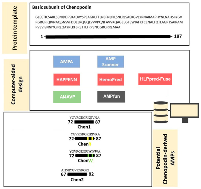
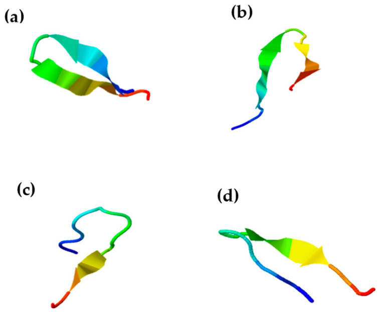
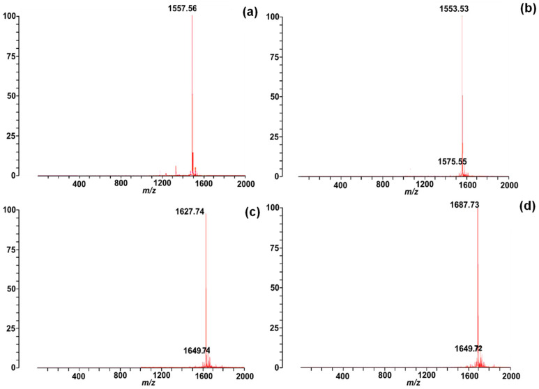
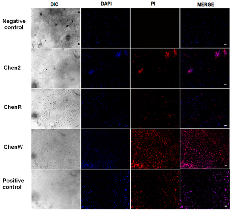
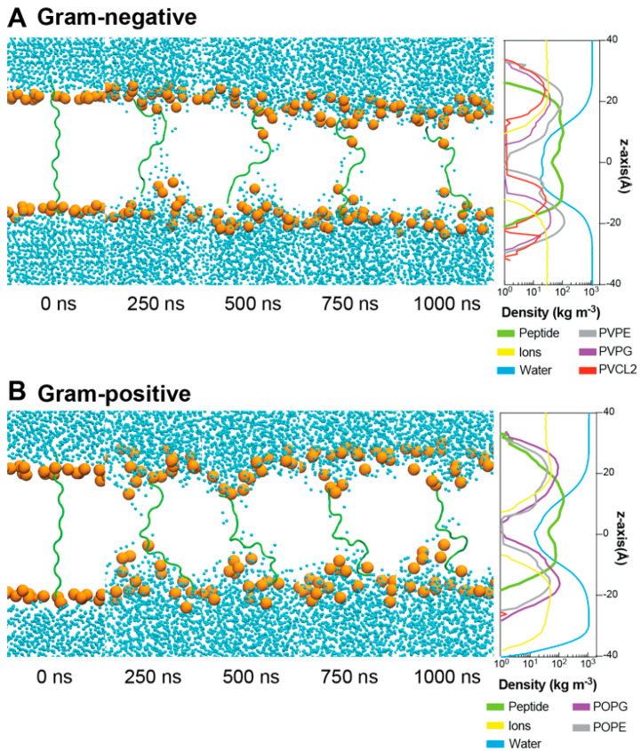
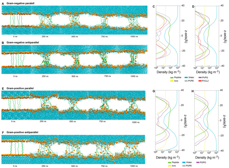
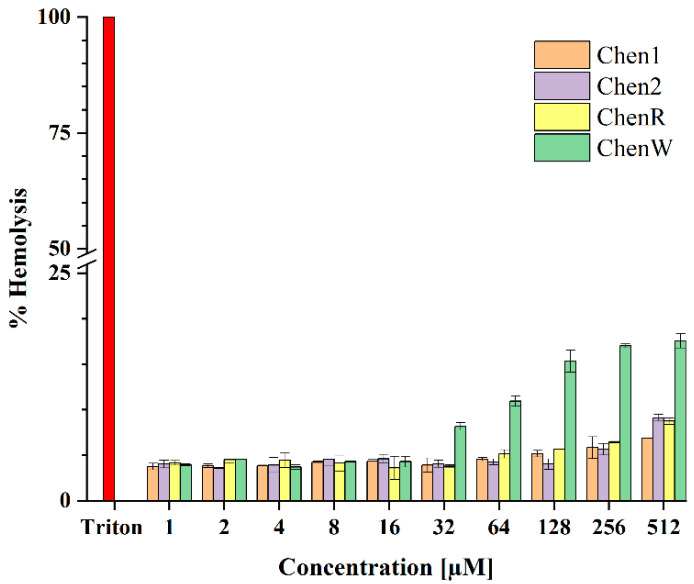
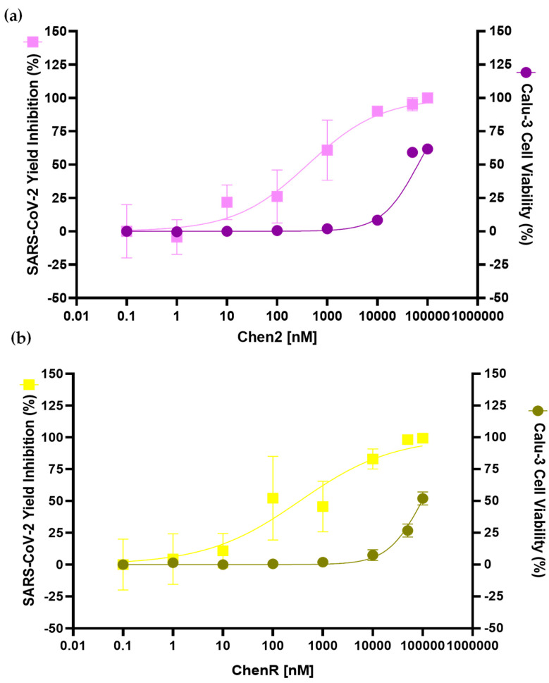
Antimicrobial peptides have been developed based on plant-derived molecular scaffolds for the treatment of infectious diseases. Chenopodin is an abundant seed storage protein in quinoa, an Andean plant with high nutritional and therapeutic properties. Here, we used computer- and physicochemical-based strategies and designed four peptides derived from the primary structure of Chenopodin. Two peptides reproduce natural fragments of 14 amino acids from Chenopodin, named Chen1 and Chen2, and two engineered peptides of the same length were designed based on the Chen1 sequence. The two amino acids of Chen1 containing amide side chains were replaced by arginine (ChenR) or tryptophan (ChenW) to generate engineered cationic and hydrophobic peptides. The evaluation of these 14-mer peptides on Staphylococcus aureus and Escherichia coli showed that Chen1 does not have antibacterial activity up to 512 µM against these strains, while other peptides exhibited antibacterial effects at lower concentrations. The chemical substitutions of glutamine and asparagine by amino acids with cationic or aromatic side chains significantly favoured their antibacterial effects. These peptides did not show significant hemolytic activity. The fluorescence microscopy analysis highlighted the membranolytic nature of Chenopodin-derived peptides. Using molecular dynamic simulations, we found that a pore is formed when multiple peptides are assembled in the membrane. Whereas, some of them form secondary structures when interacting with the membrane, allowing water translocations during the simulations. Finally, Chen2 and ChenR significantly reduced SARS-CoV-2 infection. These findings demonstrate that Chenopodin is a highly useful template for the design, engineering, and manufacturing of non-toxic, antibacterial, and antiviral peptides.

Keywords: Chenopodin; antimicrobial; antiviral; membranolytic; quinoa; synthetic peptides.


Conflict of interest statement

The authors declare no conflicts of interest.

Figures

Figure 1
Figure 1
Antimicrobial and hemolytic screening of the primary structure of the basic subunit of Chenopodin using bioinformatic tools. In silico approaches employed to predict antibacterial, hemolytic, and antiviral activities are highlighted in blue, red, and green, respectively. AMPfun is coloured in grey because it was used to predict both antibacterial and antiviral properties. Most of these sequence-based predictors classify the peptide in antimicrobial/non-antimicrobial, hemolytic/non-hemolytic and antiviral/non-antiviral or show a column “score”, which indicates how much probability a peptide candidate displays a certain effect. According to AMPA software, this protein subunit has a potential antimicrobial region (72–87 segment—Chen1). We also selected another region (67–82 segment—Chen2) with potential antibacterial and antiviral actions and low haemolytic tendency. Two new peptide analogues (ChenR and ChenW) were designed by replacing two amino acids in Chen 1. Replacement sites in the sequence are highlighted in green and yellow. Yellow dots represent substitutions with arginine, while green symbolises that amino acids have been replaced by tryptophan. Overall, cationicity-enhanced and hydrophobicity-optimised analogues showed a higher probability of exerting antibacterial effects than Chen-1. The differences between the sequences of ChenR and ChenW peptides compared to Chen1 are highlighted in bold.
Figure 2
Figure 2
Predicted secondary structures of Chenopodin-derived peptides, (a) Chen 1, (b) Chen2, (c) ChenR, and (d) ChenW. The structures were predicted using the I-TASSER platform. PED2D predictor was also employed to confirm the suggested secondary structures. The results are shown in Supplementary Material Table S7.
Figure 3
Figure 3
Mass spectra of Chenopodin-derived peptides: (a) Chen1, m/z = 1557.56 Da; (b) Chen2, m/z = 1553.53 Da; (c) ChenR, m/z = 1627.74 Da; (d) ChenW, m/z = 1687.73 Da, as identified through MALDI-ToF mass spectrometry.
Figure 4
Figure 4
Chenopodin-derived peptides affect the bacterial membrane integrity. The bacterial membrane integrity was analysed using dual staining (DAPI/PI) by fluorescence micrographs on S. aureus incubated with Chen2, ChenR, and ChenW at 1× MIC. Untreated cells served as the negative control, and 10% (v/v) SDS was used as the positive control. The scale bars are 10 μm.
Figure 5
Figure 5
Unbiased molecular dynamic simulations of ChenW peptide embedded in Gram-negative (A) and Gram-positive (B) membranes reveal spontaneous water translocation. For better representation, only phosphate groups (orange), water (blue), and peptides (green) are displayed through the 1000 ns-MDs.
Figure 6
Figure 6
Unbiased molecular dynamics simulations of multiple (six) ChenW peptides embedded in Gram-negative (A,B) and Gram-positive (E,F) membranes reveal spontaneous water translocation. Density profiles for Gram-negative membranes and ChenW peptides in parallel (C) and antiparallel (D) conformations, as well as Gram-positive membranes and peptides in parallel (G) and antiparallel (H) conformations. For better representation, only phosphate groups (orange), waters(blue), and the peptides (green) are displayed through the 1000 ns-MDs.
Figure 7
Figure 7
Evaluation of haemolytic activity of Chenopodin-derived peptides. Human red blood cells were incubated with synthetic peptides (1.0–512 µM) at 37 °C for 2 h. The level of haemolysis observed in the positive control (Triton X-100) was considered as 100% to calculate the levels in other samples. Data represent the mean ± S.D. (n = 3).
Figure 8
Figure 8
Antiviral activity of Chen2 (a) and ChenR (b) peptides in SARS-CoV-2-mediated infection of human pulmonary cells. SARS-CoV-2 yield inhibition (%), and cellular viability of Calu-3-infected cells are depicted in the graphs. Infected cells were incubated with a range of concentrations of each peptide. The SARS-CoV-2 yield inhibition (%) values were calculated in comparison to the negative control (infected, untreated cells). The cell viability assessed by MTT assay was expressed as % of mortality values and represents the means of eight repetitions with SD error bars. The SARS-CoV-2 Yield Inhibition (%) is a means of three repetitions with SD error bars.

References

    1. International Monetary Fund (IMF) World Economic Outlook: Recovery during a Pandemic-Health Concerns, Supply Disruptions, Price Pressures. International Monetary Fund; Washington, DC, USA: 2021.
    1. Baker R.E., Mahmud A.S., Miller I.F., Rajeev M., Rasambainarivo F., Rice B.L., Takahashi S., Tatem A.J., Wagner C.E., Wang L.-F., et al. Infectious Disease in an Era of Global Change. Nat. Rev. Microbiol. 2022;20:193–205. doi: 10.1038/s41579-021-00639-z. - DOI - PMC - PubMed
    1. World Health Organization (WHO) WHO Coronavirus (COVID-19) Dashboard. [(accessed on 31 December 2023)]; Available online: https://data.who.int/dashboards/covid19/cases?n=c.
    1. Lai C.-C., Chen S.-Y., Ko W.-C., Hsueh P.-R. Increased Antimicrobial Resistance during the COVID-19 Pandemic. Int. J. Antimicrob. Agents. 2021;57:106324. doi: 10.1016/j.ijantimicag.2021.106324. - DOI - PMC - PubMed
    1. Ghosh S., Bornman C., Zafer M.M. Antimicrobial Resistance Threats in the Emerging COVID-19 Pandemic: Where Do We Stand? J. Infect. Public Health. 2021;14:555–560. doi: 10.1016/j.jiph.2021.02.011. - DOI - PMC - PubMed

LinkOut - more resources