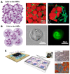
Hydrogel Microparticles for Bone Regeneration
- PMID: 38247752
- PMCID: PMC10815488
- DOI: 10.3390/gels10010028
Hydrogel Microparticles for Bone Regeneration
Abstract
Hydrogel microparticles (HMPs) stand out as promising entities in the realm of bone tissue regeneration, primarily due to their versatile capabilities in delivering cells and bioactive molecules/drugs. Their significance is underscored by distinct attributes such as injectability, biodegradability, high porosity, and mechanical tunability. These characteristics play a pivotal role in fostering vasculature formation, facilitating mineral deposition, and contributing to the overall regeneration of bone tissue. Fabricated through diverse techniques (batch emulsion, microfluidics, lithography, and electrohydrodynamic spraying), HMPs exhibit multifunctionality, serving as vehicles for drug and cell delivery, providing structural scaffolding, and functioning as bioinks for advanced 3D-printing applications. Distinguishing themselves from other scaffolds like bulk hydrogels, cryogels, foams, meshes, and fibers, HMPs provide a higher surface-area-to-volume ratio, promoting improved interactions with the surrounding tissues and facilitating the efficient delivery of cells and bioactive molecules. Notably, their minimally invasive injectability and modular properties, offering various designs and configurations, contribute to their attractiveness for biomedical applications. This comprehensive review aims to delve into the progressive advancements in HMPs, specifically for bone regeneration. The exploration encompasses synthesis and functionalization techniques, providing an understanding of their diverse applications, as documented in the existing literature. The overarching goal is to shed light on the advantages and potential of HMPs within the field of engineering bone tissue.
Keywords: HMP-based scaffolds; HMP-incorporated scaffolds; bioactive-factor delivery; bone regeneration; cell delivery; hydrogel microparticles; hydrogels; microgels; osteogenesis; tissue engineering; tissue scaffolds.
Conflict of interest statement
The authors declare no conflict of interest.
Figures


Similar articles
-
Hydrogel microparticles for biomedical applications.Nat Rev Mater. 2020 Jan;5(1):20-43. doi: 10.1038/s41578-019-0148-6. Epub 2019 Nov 7. Nat Rev Mater. 2020. PMID: 34123409 Free PMC article.
-
Enhanced in vivo retention of low dose BMP-2 via heparin microparticle delivery does not accelerate bone healing in a critically sized femoral defect.Acta Biomater. 2017 Sep 1;59:21-32. doi: 10.1016/j.actbio.2017.06.028. Epub 2017 Jun 20. Acta Biomater. 2017. PMID: 28645809 Free PMC article.
-
Granular hydrogels: emergent properties of jammed hydrogel microparticles and their applications in tissue repair and regeneration.Curr Opin Biotechnol. 2019 Dec;60:1-8. doi: 10.1016/j.copbio.2018.11.001. Epub 2018 Nov 24. Curr Opin Biotechnol. 2019. PMID: 30481603 Free PMC article. Review.
-
Microgels: Modular, tunable constructs for tissue regeneration.Acta Biomater. 2019 Apr 1;88:32-41. doi: 10.1016/j.actbio.2019.02.011. Epub 2019 Feb 12. Acta Biomater. 2019. PMID: 30769137 Free PMC article. Review.
-
Advancing bioinks for 3D bioprinting using reactive fillers: A review.Acta Biomater. 2020 Sep 1;113:1-22. doi: 10.1016/j.actbio.2020.06.040. Epub 2020 Jul 2. Acta Biomater. 2020. PMID: 32622053 Review.
Cited by
-
Dual Delivery of Cells and Bioactive Molecules for Wound Healing Applications.Molecules. 2025 Mar 31;30(7):1577. doi: 10.3390/molecules30071577. Molecules. 2025. PMID: 40286165 Free PMC article.
-
Optimized Adipogenic Differentiation and Delivery of Bovine Umbilical Cord Stem Cells for Cultivated Meat.Gels. 2024 Jul 24;10(8):488. doi: 10.3390/gels10080488. Gels. 2024. PMID: 39195017 Free PMC article.
-
Hydrogel-Based Scaffolds: Advancing Bone Regeneration Through Tissue Engineering.Gels. 2025 Feb 27;11(3):175. doi: 10.3390/gels11030175. Gels. 2025. PMID: 40136878 Free PMC article. Review.
-
Human Mesenchymal Stem Cells on Size-Sorted Gelatin Hydrogel Microparticles Show Enhanced In Vitro Wound Healing Activities.Gels. 2024 Jan 26;10(2):97. doi: 10.3390/gels10020097. Gels. 2024. PMID: 38391427 Free PMC article.
-
Expansion and Delivery of Human Chondrocytes on Gelatin-Based Cell Carriers.Gels. 2025 Mar 13;11(3):199. doi: 10.3390/gels11030199. Gels. 2025. PMID: 40136904 Free PMC article.
References
Publication types
LinkOut - more resources
Full Text Sources

