Review
. 2024 Jan 6;10(1):45.
doi: 10.3390/gels10010045.

Novel Therapeutic Hybrid Systems Using Hydrogels and Nanotechnology: A Focus on Nanoemulgels for the Treatment of Skin Diseases

Affiliations
Review

Novel Therapeutic Hybrid Systems Using Hydrogels and Nanotechnology: A Focus on Nanoemulgels for the Treatment of Skin Diseases

Kamil Sghier et al. Gels. .

Abstract

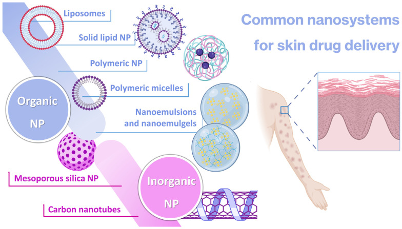
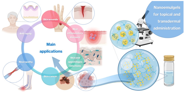
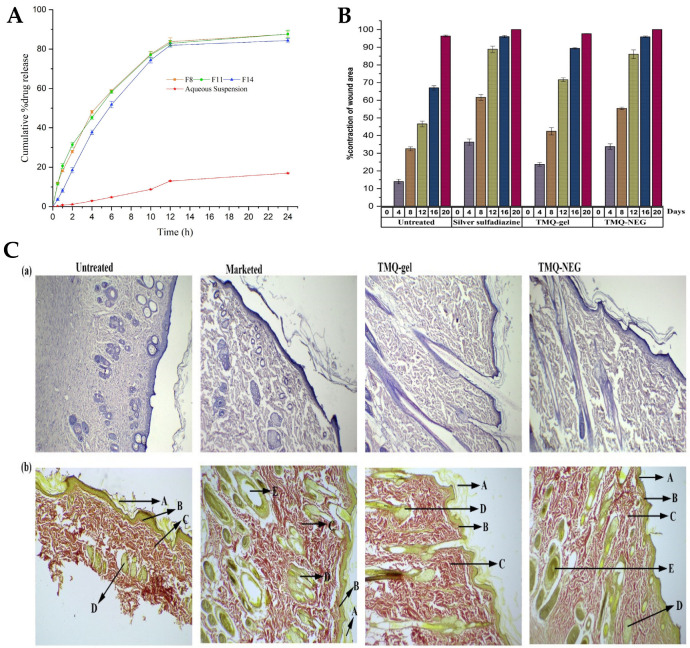
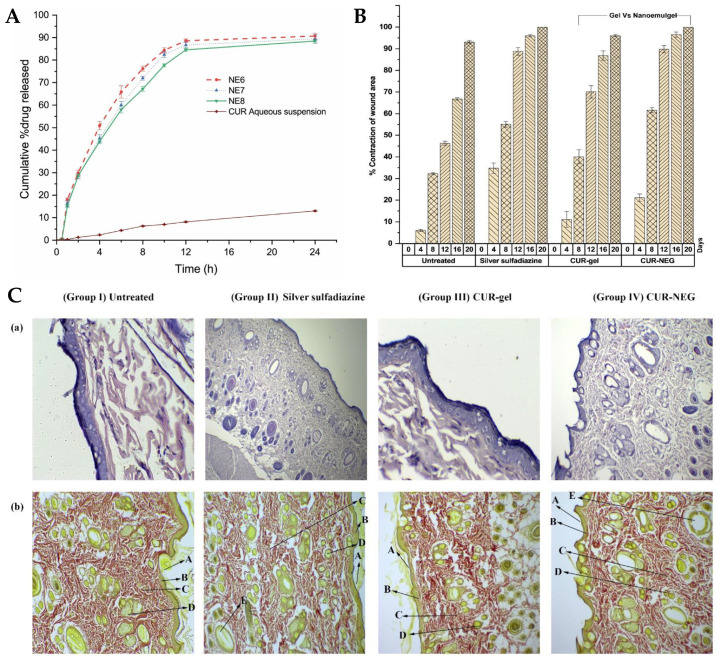
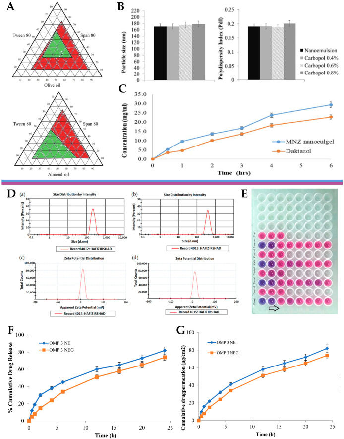
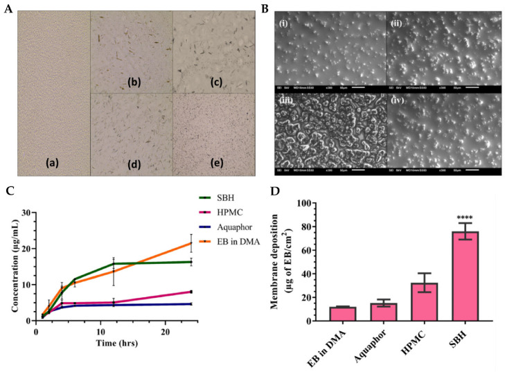
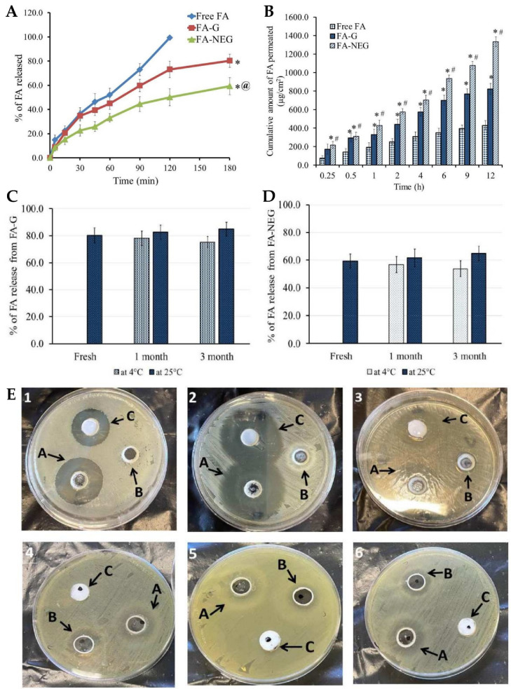
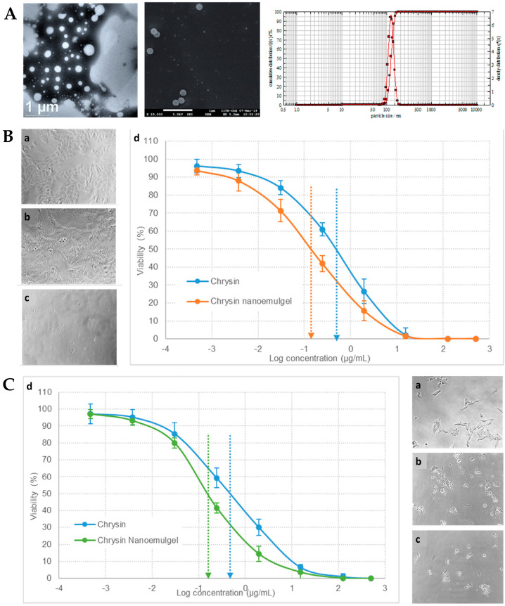
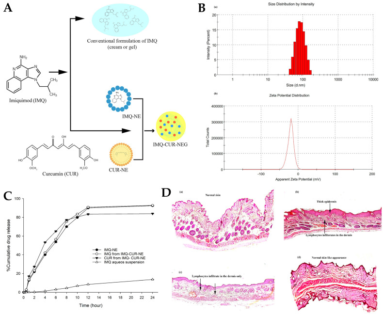
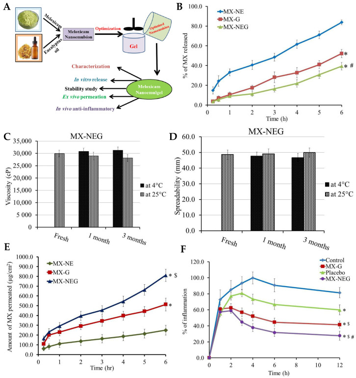
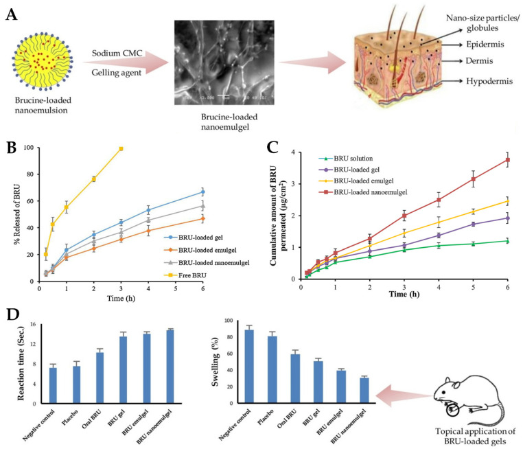
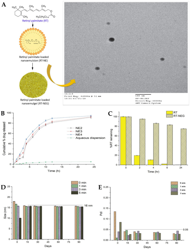
Topical and transdermal drug delivery are advantageous administration routes, especially when treating diseases and conditions with a skin etiology. Nevertheless, conventional dosage forms often lead to low therapeutic efficacy, safety issues, and patient noncompliance. To tackle these issues, novel topical and transdermal platforms involving nanotechnology have been developed. This review focuses on the latest advances regarding the development of nanoemulgels for skin application, encapsulating a wide variety of molecules, including already marketed drugs (miconazole, ketoconazole, fusidic acid, imiquimod, meloxicam), repurposed marketed drugs (atorvastatin, omeprazole, leflunomide), natural-derived compounds (eucalyptol, naringenin, thymoquinone, curcumin, chrysin, brucine, capsaicin), and other synthetic molecules (ebselen, tocotrienols, retinyl palmitate), for wound healing, skin and skin appendage infections, skin inflammatory diseases, skin cancer, neuropathy, or anti-aging purposes. Developed formulations revealed adequate droplet size, PDI, viscosity, spreadability, pH, stability, drug release, and drug permeation and/or retention capacity, having more advantageous characteristics than current marketed formulations. In vitro and/or in vivo studies established the safety and efficacy of the developed formulations, confirming their therapeutic potential, and making them promising platforms for the replacement of current therapies, or as possible adjuvant treatments, which might someday effectively reach the market to help fight highly incident skin or systemic diseases and conditions.

Keywords: anti-aging; nanoemulgels; nanoemulsions; neuropathy; skin cancer; skin infection; skin inflammation; topical administration; transdermal administration; wound healing.


Conflict of interest statement

The authors declare no conflicts of interest.

Figures

Figure 1
Figure 1
Schematic representation of common nanosystem categories for skin drug delivery (produced with Biorender).
Figure 2
Figure 2
Schematic representation of nanoemulgel structure, and main applications of nanoemulgels in highly incident diseases, for topical and transdermal administration (produced with Biorender).
Figure 3
Figure 3
(A) Droplet size distribution of the developed ATR nanoemulgel; (B) surface morphology of the developed ATR nanoemulgel; (C) in vitro drug release profiles of the developed ATR nanoemulgel, compared to an emulgel and gel; (D) ex vivo drug permeation profiles of the developed ATR nanoemulgel, compared to an emulgel, a gel, and a solution; (E) wound area variation of rat skin after topical administration of the developed ATR formulations, after 0, 7, 14, and 21 treatment days; (F,G)—healing score (F) and photomicrographs (G) of rat skin before treatment (a), and after 21 days of topical administration of an ATR gel (b), an ATR emulgel (c), or an ATR nanoemulgel (d), where black arrows represent the absence of epidermal layer epithelization, blue arrows represent loss of collagen fibers normal arrangement in the dermal layer, green arrows represent severe congestion, yellow arrows represent hemorrhage, and black circles represent inflammatory cell infiltrations; ATR—atorvastatin; adapted from Morsy et al. [149], reproduced with permission from MDPI (Creative Commons CC BY 4.0 470 license).
Figure 4
Figure 4
(A) Schematic representation of the developed eucalyptus oil nanoemulgel, including partial composition and general indication of performed studies; (B) in vitro eucalyptol release profiles of different nanoemulgels; adapted from Rehman et al. [150], reproduced with permission from MDPI (Creative Commons CC BY 4.0 470 license).
Figure 5
Figure 5
(A) In vitro drug release profiles of the TMQ-loaded nanoemulsions, compared to a drug aqueous suspension; (B) percentage of contraction of wound area variation, in the in vivo study, assessed for 20 days, with topical application of the developed TMQ nanoemulgel (TMQ-NEG), a conventional TMQ gel (TMQ-gel), a silver sulfadiazine formulation (Silver sulfadiazine), or no treatment (Untreated); (C) histopathology analysis of the rat’s skin at day 20, newly healed, after topical application of the developed TMQ nanoemulgel (TMQ-NEG), a conventional TMQ gel (TMQ-gel), a silver sulfadiazine formulation (Marketed), or no treatment (Untreated), stained with hematoxylin-eosin (a) or Van Gieson (b), with arrows indicating the stratum corneum (A), the papillary dermis (B), collagen fibers (C), sebaceous glands (D), or hair follicles (E); adapted from Algahtani et al. [152], reproduced with permission from MDPI (Creative Commons CC BY 4.0 470 license).
Figure 6
Figure 6
(A) In vitro cumulative drug release from the developed preliminary curcumin nanoemulsions, compared to the control (drug suspension); (B) in vivo wound-healing activity, in a rat model, of the developed curcumin nanoemulgel (CUR-NEG), compared to a curcumin conventional gel (CUR-gel), a marketed control formulation (Silver sulfadiazine), or no treatment (Untreated), including contraction of wound area percentage; (C) histopathology analysis of the rat’s skin tissue at day 20 after treatment, including indications for the stratum corneum (A), the papillary dermis (B), collagen fibers (C), sebaceous glands (D), and hair follicles (E), (a) Stained with hematoxylin-eosin; (b) stained with vangeison to observe collagen formation (at 10× magnification); adapted from Algahtani et al. [153], reproduced with permission from MDPI (Creative Commons CC BY 4.0 470 license).
Figure 7
Figure 7
(A) Ternary phase diagrams of the preliminary nanoemulsions containing either olive oil, Tween® 80, and Span® 80, or almond oil, Tween® 80, and Span® 80; (B) droplet size and polydispersity index of the developed miconazole nitrate preliminary nanoemulsion and nanoemulgel formulations; (C) in vitro drug release profiles, in Franz diffusion cells, of the developed miconazole nitrate nanoemulgel, compared to the marketed product, Daktazol® cream; adapted from Tayah et al. [154], reproduced with permission from Elsevier (license number 5671991093263); (D) droplet size (a) and zeta potential (c) of the developed omeprazole-loaded nanoemulsion, and droplet size (b) and zeta potential (d) of the developed omeprazole-loaded nanoemulgel; (E) minimum inhibitory concentration determination assay of the developed omeprazole-loaded nanoemulgel, against selected bacterial strains, using a 96-well microplate (arrow shows decrescent antimicrobial activity); (F) cumulative drug release percentage from the developed omeprazole-loaded nanoemulsion and nanoemulgel formulations; (G) cumulative drug permeation from the developed omeprazole-loaded nanoemulsion and nanoemulgel formulations; adapted from Ullah et al. [155], reproduced with permission from MDPI (Creative Commons CC BY 4.0 470 license).
Figure 8
Figure 8
(A) Optical microscopy images of the EB-loaded preliminary nanoemulgels, containing either Soluplus® (a), HPMC (b), Poloxamer 407 (c), Carbopol® 974P (d), or Aquaphor (e); (B) scanning electron microscopy images of the optimized nanoemulgels, either drug-loaded Soluplus® formulation (i), Soluplus® vehicle (ii), drug-loaded HPMC formulation (iii), or HPMC vehicle (iv); (C,D) in vitro cumulative drug release (C) and membrane drug deposition (D) of the different EB-loaded nanoemulgels; **** p < 0.0001; DMA—dimethylacetamide; EB—Ebselen; HPMC—hydroxypropyl methylcellulose; SBH—Soluplus®; adapted from Vartak et al. [156], reproduced with permission from Elsevier (license number 5672000024708).
Figure 9
Figure 9
(A) In vitro FA release profiles from the developed nanoemulgel (FA-NEG), compared to a conventional gel (FA-G) and a drug suspension (Free FA), where * p < 0.05 compared to the drug suspension, and @ p < 0.05 compared to the conventional gel; (B) ex vivo FA permeation profiles, across rat skin, from the developed nanoemulgel (FA-NEG), compared to a conventional gel (FA-G) and a drug suspension (Free FA), where * p < 0.05 compared to the drug suspension, and # p < 0.05 compared to the conventional gel; (C,D) variation of the in vitro drug release during stability studies, under storage at 4 °C and 25 °C for 1 and 3 months, for the developed nanoemulgel (C) and conventional gel (D) formulations; (E) inhibition zone diameter photographs after treatment with the developed FA-loaded nanoemulgel (A), placebo nanoemulgel (B), or marketed FA formulation (C), on Bacillus subtilis (1), Staphylococcus aureus (2), Enterococcus faecalis (3), Candida albicans (4), Shigella (5), and Escherichia coli (6); FA—fusidic acid; adapted from Almostafa et al. [158], reproduced with permission from MDPI (Creative Commons CC BY 4.0 470 license).
Figure 10
Figure 10
(A) Developed chrysin nanoformulation’s droplet size and size distribution, with transmission electron microscopy image (left), scanning electron microscopy image (middle), and photon cross-correlation spectroscopy results (right); (B) A375 cells’ morphological observation and growth inhibition after no treatment (control cells, a), treatment with chrysin solution (b), or treatment with the developed chrysin nanoemulgel (c), and respective in vitro cytotoxicity profile (cell viability %) (d); (C) SK-MEL-2 cells’ morphological observation and growth inhibition after no treatment (control cells, a), treatment with chrysin solution (b), or treatment with the developed chrysin nanoemulgel (c), and respective in vitro cytotoxicity profile (cell viability %) (d); adapted from Nagaraja et al. [142], reproduced with permission from MDPI (Creative Commons CC BY 4.0 470 license).
Figure 11
Figure 11
(A) Schematic representation of the developed IMQ-CUR-nanoemulgel; (B) droplet size (a) and zeta potential (b) distribution of the developed preliminary nanoemulsion; (C) in vitro IMQ and CUR drug release profiles from different formulations, including an IMQ-nanoemulsion (IMQ-NE, with no CUR), IMQ-CUR-nanoemulsion (with both IMQ and CUR), and an IMQ aqueous suspension (control); (D) histopathology images of the mice’s skin after the ten days of topical treatment, with either the IMQ gel (b), the IMQ-nanoemulgel (c), the IMQ-CUR-nanoemulgel (d), or no treatment (a); CUR—curcumin; IMQ—imiquimod; adapted from Algahtani et al. [159], reproduced with permission from MDPI (Creative Commons CC BY 4.0 470 license).
Figure 12
Figure 12
(A) Schematic representation of the developed MX and eucalyptus oil nanoemulgel, including performed physicochemical and efficacy characterization studies; (B) in vitro drug release profiles of the developed preliminary MX-nanoemulsion (MX-NE), conventional MX-gel (MX-G), and MX-nanoemulgel (MX-NEG), with * p < 0.05 compared to the preliminary MX-nanoemulsion, and # p < 0.05 compared to the conventional MX-gel; (C,D) stability profiles of the developed MX-nanoemulgel formulation, after 1 and 3 months, under storage at 4 °C and 25 °C, in what concerns viscosity (C) and spreadability (D); (E) ex vivo drug permeation profiles of the developed preliminary MX-nanoemulsion (MX-NE), conventional MX-gel (M-G), and MX-nanoemulgel (MG-NEG), with * p < 0.05 compared to the preliminary MX-nanoemulsion, and $ p < 0.05 compared to the conventional MX-gel; (F) anti-inflammatory effects of various formulations on rat hind-paw edema, including the developed MX-nanoemulgel (MG-NEG), a conventional MX-gel (M-G), a placebo formulation (nanoemulgel vehicle with no drug), and a control group (no treatment), with * p < 0.05 compared to the control group, $ p < 0.05 compared to the placebo group, and # p < 0.05 compared to the conventional MX-gel group; MX—meloxicam; adapted from Shehata et al. [160], reproduced with permission from MDPI (Creative Commons CC BY 4.0 470 license).
Figure 13
Figure 13
(A) Schematic representation of the developed brucine-loaded topical nanoemulgel, including a scanning electron microscopy image; (B) in vitro drug release profiles from different BRU-loaded formulations, namely, a drug solution (Free BRU), a conventional gel, an emulgel, and a nanoemulgel; (C) ex vivo drug permeation profiles from different BRU-loaded formulations, namely, a drug solution, a conventional gel, an emulgel, and a nanoemulgel; (D) antinociceptive (reaction time) and anti-inflammatory (swelling) effects of different brucine-loaded formulations after administration to mice; BRU—brucine; adapted from Abdallah et al. [161], reproduced with permission from Elsevier (license number 5672000458127).
Figure 14
Figure 14
(A) Schematic representation of the developed retinyl palmitate topical nanoemulgel (left), with corresponding transmission electron microscopy image showing droplet morphology and size (right); (B) in vitro drug release profiles of different retinyl palmitate-loaded preliminary nanoemulsions, compared to an aqueous dispersion of the drug; (C) UV stability of the developed retinyl palmitate nanoemulgel, compared to the nonencapsulated drug; (D) mean droplet size variations of the preliminary optimized retinyl palmitate-loaded nanoemulsions as effect of storage time; (E) PDI variations of the preliminary optimized retinyl palmitate-loaded nanoemulsions as effect of storage time; NE—nanoemulsion; NEG—nanoemulgel; PDI—polydispersity index; RT—retinyl palmitate; adapted from Algahtani et al. [163], reproduced with permission from MDPI (Creative Commons CC BY 4.0 470 license).

Similar articles

Cited by

References

    1. Arda O., Göksügür N., Tüzün Y. Basic Histological Structure and Functions of Facial Skin. Clin. Dermatol. 2014;32:3–13. doi: 10.1016/j.clindermatol.2013.05.021. - DOI - PubMed
    1. Boer M., Duchnik E., Maleszka R., Marchlewicz M. Structural and Biophysical Characteristics of Human Skin in Maintaining Proper Epidermal Barrier Function. Adv. Dermatol. Allergol. 2016;1:1–5. doi: 10.5114/pdia.2015.48037. - DOI - PMC - PubMed
    1. Abdo J.M., Sopko N.A., Milner S.M. The Applied Anatomy of Human Skin: A Model for Regeneration. Wound Med. 2020;28:100179. doi: 10.1016/j.wndm.2020.100179. - DOI
    1. Baroni A., Buommino E., De Gregorio V., Ruocco E., Ruocco V., Wolf R. Structure and Function of the Epidermis Related to Barrier Properties. Clin. Dermatol. 2012;30:257–262. doi: 10.1016/j.clindermatol.2011.08.007. - DOI - PubMed
    1. Xie J., Hao T., Li C., Wang X., Yu X., Liu L. Automatic Evaluation of Stratum Basale and Dermal Papillae Using Ultrahigh Resolution Optical Coherence Tomography. Biomed. Signal Process Control. 2019;53:101527. doi: 10.1016/j.bspc.2019.04.004. - DOI

LinkOut - more resources