Review
. 2024 Jan 12;13(2):150.
doi: 10.3390/cells13020150.

Involvement of Astrocytes in the Formation, Maintenance, and Function of the Blood-Brain Barrier

Affiliations
Review

Involvement of Astrocytes in the Formation, Maintenance, and Function of the Blood-Brain Barrier

Gabriella Schiera et al. Cells. .

Abstract

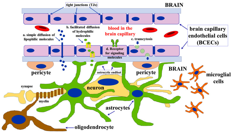
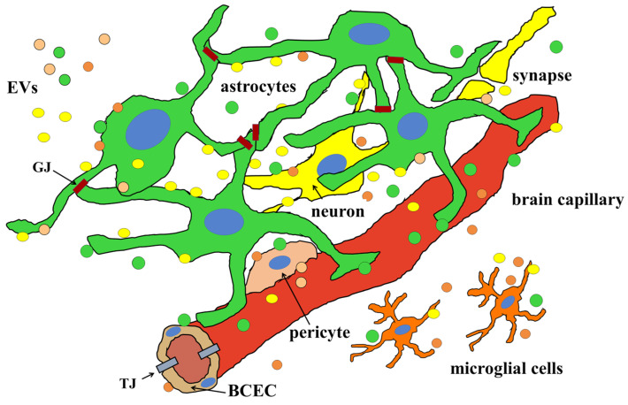
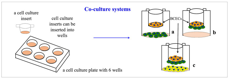
The blood-brain barrier (BBB) is a fundamental structure that protects the composition of the brain by determining which ions, metabolites, and nutrients are allowed to enter the brain from the blood or to leave it towards the circulation. The BBB is structurally composed of a layer of brain capillary endothelial cells (BCECs) bound to each other through tight junctions (TJs). However, its development as well as maintenance and properties are controlled by the other brain cells that contact the BCECs: pericytes, glial cells, and even neurons themselves. Astrocytes seem, in particular, to have a very important role in determining and controlling most properties of the BBB. Here, we will focus on these latter cells, since the comprehension of their roles in brain physiology has been continuously expanding, even including the ability to participate in neurotransmission and in complex functions such as learning and memory. Accordingly, pathological conditions that alter astrocytic functions can alter the BBB's integrity, thus compromising many brain activities. In this review, we will also refer to different kinds of in vitro BBB models used to study the BBB's properties, evidencing its modifications under pathological conditions.

Keywords: astrocytes; blood–brain barrier; brain capillary endothelial cell; extracellular vesicles (EVs); in vitro BBB models.


Conflict of interest statement

The authors declare no conflict of interest.

Figures

Figure 1
Figure 1
Schematic view of all the components of the neurovascular unit (NVU). The brain capillary is structurally composed of the brain capillary endothelial cells (BCECs) bound together by tight junctions (TJs). The formation of the blood–brain barrier depends, however, on all the cells present around the capillary: neurons (yellow in the picture), oligodendrocytes (brown in the figure), microglial cells (orange in the picture), pericytes (light red in the picture), and, especially, astrocytes (green in the picture). All these cells communicate with each other and with the BCECs by releasing soluble factors that, as discussed below, can be also conveyed by extracellular vesicles. Astrocytes, in particular, directly contact BCECs through the so-called astrocytic endfeet, which also contains aquaporins (see the text for further details).
Figure 2
Figure 2
All the components of the neurovascular unit (NVU) are able to release and receive extracellular vesicles (EVs). As discussed in the text, the walls of the brain capillaries are structurally composed of the BCECs bound together by tight junctions (TJs); the formation of a functional blood–brain barrier depends, however, on all the cells present around the capillary. All these cells communicate with each other both through direct contacts and secreted molecules, many of which are delivered by the EVs. For clarity, EVs coming from different cell types have been represented in the same color as the producing cells. Notably, astrocytes form a sort of large web thanks to their ability to form gap junctions (GJs) with each other.
Figure 3
Figure 3
Simple co-culture systems (also called transwell systems) used for studying the role of different brain cell types on BBB formation. This system is based on culture plates with wells that can also host inserts with a porous membrane at their bottom. Endothelial cells (orange in the figure) are plated inside the insert, on the porous membrane, enriched with proteins of the basement membrane existing in vivo. Other brain cells (green in the figure) can be cultured either at the bottom of the wells (a) or on the outside of the insert, that is on the other side of the porous membrane (b). This system can also be used for studying synergistic effects on the barrier formation of different brain cell types (for example, neurons and astrocytes, or pericytes and astrocytes). These cells can be cultured as a mixed population or separated by culturing one cell type at the bottom of the wells (yellow cells in the figure) and on the outside of the inserts of the other one (green cells in the figure) (c).

References

    1. Ballabh P., Braun A., Nedergaard M. The blood-brain barrier: An overview: Structure, regulation, and clinical implications. Neurobiol. Dis. 2004;16:1–13. doi: 10.1016/j.nbd.2003.12.016. - DOI - PubMed
    1. Abbott N.J., Rönnbäck L., Hansson E. Astrocyte-endothelial interactions at the blood-brain barrier. Nat. Rev. Neurosci. 2006;7:41–53. doi: 10.1038/nrn1824. - DOI - PubMed
    1. Serlin Y., Shelef I., Knyazer B., Friedman A. Anatomy and physiology of the blood-brain barrier. Semin. Cell Dev. Biol. 2015;38:2–6. doi: 10.1016/j.semcdb.2015.01.002. - DOI - PMC - PubMed
    1. Harder D.R., Zhang C., Gebremedhin D. Astrocytes function in matching blood flow to metabolic activity. News Physiol. Sci. 2002;17:27–31. doi: 10.1152/physiologyonline.2002.17.1.27. - DOI - PubMed
    1. Blanchette M., Daneman R. Formation and maintenance of the BBB. Mech. Dev. 2015;138:8–16. doi: 10.1016/j.mod.2015.07.007. - DOI - PubMed

LinkOut - more resources