Review
. 2023 Dec 26;11(1):28.
doi: 10.3390/bioengineering11010028.

Salivary Gland Bioengineering

Affiliations
Review

Salivary Gland Bioengineering

Stephen C Rose et al. Bioengineering (Basel). .

Abstract

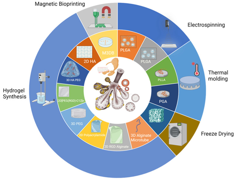
Salivary gland dysfunction affects millions globally, and tissue engineering may provide a promising therapeutic avenue. This review delves into the current state of salivary gland tissue engineering research, starting with a study of normal salivary gland development and function. It discusses the impact of fibrosis and cellular senescence on salivary gland pathologies. A diverse range of cells suitable for tissue engineering including cell lines, primary salivary gland cells, and stem cells are examined. Moreover, the paper explores various supportive biomaterials and scaffold fabrication methodologies that enhance salivary gland cell survival, differentiation, and engraftment. Innovative engineering strategies for the improvement of vascularization, innervation, and engraftment of engineered salivary gland tissue, including bioprinting, microfluidic hydrogels, mesh electronics, and nanoparticles, are also evaluated. This review underscores the promising potential of this research field for the treatment of salivary gland dysfunction and suggests directions for future exploration.

Keywords: organoid; regeneration; salivary gland; tissue engineering.


Conflict of interest statement

The authors declare no conflicts of interest.

Figures

Figure 4
Figure 4
Myofibroblast origins and fate in normal wound healing. Created with Biorender.com [107].
Figure 9
Figure 9
Electronic mesh schematic (created using Biorender, Biorender.com) [309].
Figure 1
Figure 1
Primary salivary glands. There are three pairs of major salivary glands—parotid, submandibular, and sublingual—innervated by the facial and glossopharyngeal nerves. Created with BioRender.com (2023).
Figure 2
Figure 2
The adenomere functional unit of the human salivary gland. Reprinted with permission from de Paula et al., 2017. © 2017 Wiley Periodicals [13].
Figure 3
Figure 3
Mouse and human salivary glands develop through the process of branching morphogenesis. (a) Schematic stages of mouse salivary gland development. (b) Histological representations of mouse and human saliavry gland development. (Top panel) Mouse development measured in days. E = embryonic day. (Bottom panel) Human development measured in weeks. Reprinted with permission from Mattingly et al., 2015. © 2015 Wiley Periodicals [22].
Figure 5
Figure 5
Major approaches to biofabrication of salivary gland tissues (Created using Biorender, Biorender.com).
Figure 6
Figure 6
Nanofiber scaffolds promote self-organization and branching morphogenesis of dissociated embryonic salivary gland cells. (A) Bright field images of spontaneously re-aggregated cell pellets from dissociated embryonic day 13 (E13) salivary gland cells cultured on polycarbonate filter membrane, PLGA nanofibers, and microfibers for 48 h. Dashed lines outline the morphology of re-aggregated structures on microfibers to compensate for the interference with light microscopy. (B) Confocal images through the equatorial section of re-aggregated buds immunostaining for talin (green, top panels) or vinculin (cyan, bottom panels), co-stained for nuclei (DAPI, blue), showed diffuse cortical expression with stronger staining along the basal cell membranes at the bud periphery (arrowheads), similar to that observed in intact glands, scale bars = 50 μm. Reprinted with permission from Sequeira et al., 2012. © 2012 Elsevier [235].
Figure 7
Figure 7
Biomaterials can control shape of organized tissue constructs. (A) Confocal image (right) of SIMS cells stained for F-actin (green, phalloidin) and nuclei (blue, DAPI) grown in 30 μm nanofibrous craters (SEM of top view, left and angled view, middle) for 96 h, scale bar = 10 μm. The Z-plane is denoted by the arrow. Reprinted with permission from Soscia et al., 2013. © 2022 Elsevier [234]. (B) Confocal images of CellTracker™ Red CMTPX-labeled SIMS cells and CellTracker™ Green CMFDA-labeled NIH 3T3 fibroblasts showed cellular organization after co-cultured in microtubes for 4 days, scale bar = 250 µm. Reprinted and modified with permission from Jorgensen et al., 2022. © 2022 by the authors [244].
Figure 8
Figure 8
Cell behavior in organoids can be achieved through cell surface modifications of nanofibrous scaffolds. Confocal images of immunostained SIMS cells seeded on PLGA nanofibers (Nano) showed apical restriction of ZO-1 (green), which was disrupted by chitosan-modified nanofibers (Nano + CS) compared to 2D culture on glass or PLGA film. Scale bar = 25 µm. Reprinted with permission from Cantara et al., 2012. © 2012 Elsevier [263].

Similar articles

Cited by

References

    1. Sasportas M., Hosford A., Sodini M., Waters J., Zambricki A., Barral J., Graves E., Brinton T., Yock P., Quyhh-Thu L., et al. Cost-Effectiveness Landscape Analysis of Treatments Addressing Xerostomia in Patients Receiving Head and Neck Radiation Therapy. Oral Surg. Oral Med. Oral Pathol. Oral Raiol. 2013;116:e37–e51. doi: 10.1016/j.oooo.2013.02.017. - DOI - PMC - PubMed
    1. Turner M., Ship J. Dry Mouth and Its Effects on the Oral Health of Elderly People. J. Am. Dent. Assoc. 2007;138:S15–S20. doi: 10.14219/jada.archive.2007.0358. - DOI - PubMed
    1. Jensen S.B., Pedersen A.M.L., Vissink A., Andersen E., Brown C.G., Davies A.N., Dutilh J., Fulton J.S., Jankovic L., Lopes N.N.F., et al. A Systematic Review of Salivary Gland Hypofunction and Xerostomia Induced by Cancer Therapies: Prevalence, Severity and Impact on Quality of Life. Support. Care Cancer. 2010;18:1039–1060. doi: 10.1007/s00520-010-0827-8. - DOI - PubMed
    1. Stewart B.W., Wild C.P. World Cancer Report 2014. World Health Organization; Geneva, Switzerland: 2014.
    1. Polaris Market Research Xerostomia Therapeutics Market Share, Size, Trends, Industry Analysis Report, By Type (OTC, Prescription), By Product, By Region, And Segment Forecasts, 2023–2032, Report Summary. [(accessed on 25 August 2023)]. Available online: https://www.polarismarketresearch.com/industry-analysis/xerostomia-thera....

LinkOut - more resources