

Control of Tibial Advancement by the Plantar Flexors during the Stance Phase of Gait Depends on Knee Flexion with Respect to the Ground Reaction Force
- PMID: 38247918
- PMCID: PMC10813783
- DOI: 10.3390/bioengineering11010041
Control of Tibial Advancement by the Plantar Flexors during the Stance Phase of Gait Depends on Knee Flexion with Respect to the Ground Reaction Force
Abstract
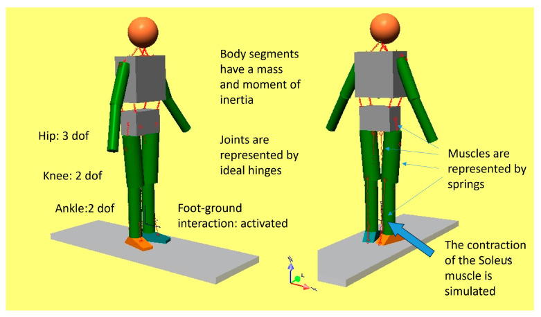
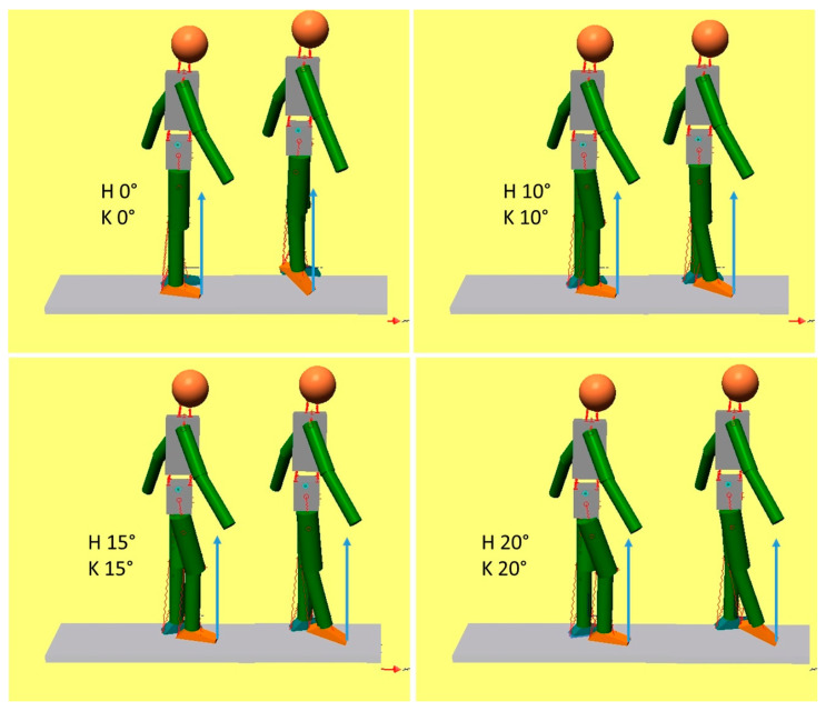
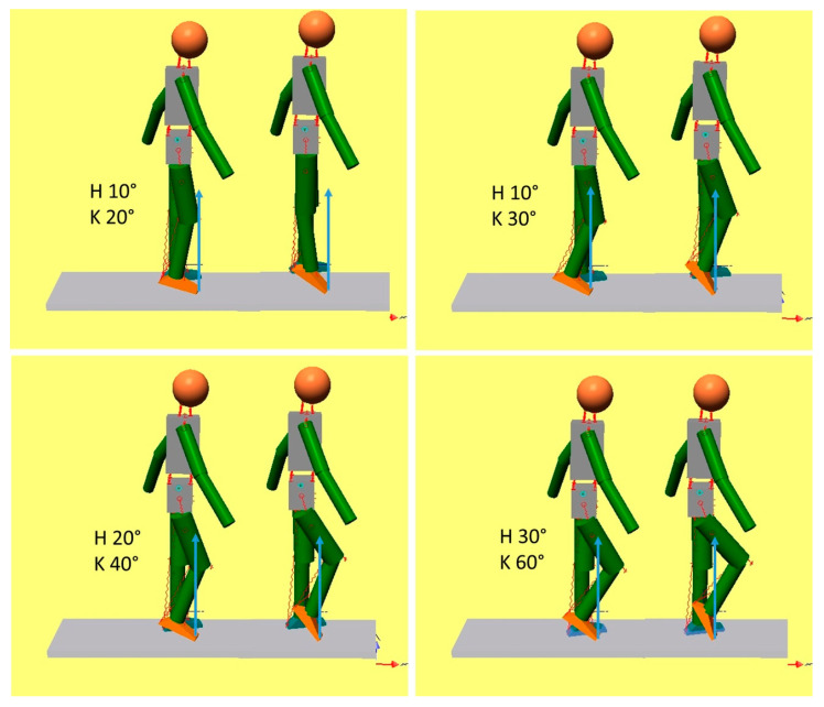
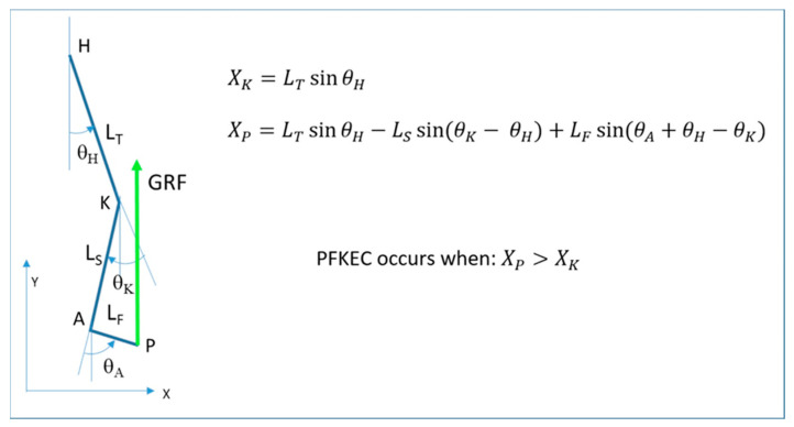
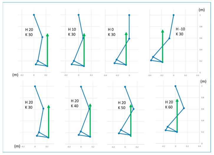
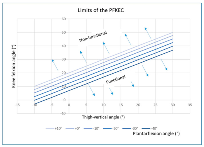
During the stance phase of a normal gait, the triceps surae muscle controls the advancement of the tibia, which contributes to knee extension. Plantar flexor weakness results in excessive dorsiflexion, and consequently, the knee loses this contribution. However, increasing knee flexion is also seen in patients with cerebral palsy who do not have plantar flexor weakness. We aimed to understand this mechanism through the use of a musculoskeletal dynamic model. The model consists of solid segments connected with rotatory joints and springs to represent individual muscles. It was positioned at different degrees of ankle plantarflexion, knee flexion, and hip flexion. The soleus muscle was activated concentrically to produce plantarflexion and push the foot against the ground. The resulting knee extension was analyzed. The principal determinant of knee flexion or extension associated with ankle plantarflexion was the position of the knee joint center. When this was anterior to the line of action of the ground reaction force (GRF), the soleus contraction resulted in increased knee flexion. The knee extension was obtained when the knee was flexed less than approximately 25°. The relation between joint angles, anthropometric parameters, and the position of the GRF was expressed in a mathematical formulation. The clinical relevance of this model is that it explains the failure of plantar flexor control on knee extension in patients with cerebral palsy, when increased knee flexion can occur even if there is a normal or plantarflexed foot position.
Keywords: crouch gait; dynamic simulation; knee flexion in gait; plantar flexion-knee extension couple; plantar flexor control on tibial advancement.
Conflict of interest statement
The authors declare no conflicts of interest.
Figures
Similar articles
-
Contributions to the understanding of gait control.Dan Med J. 2014 Apr;61(4):B4823. Dan Med J. 2014. PMID: 24814597 Review.
-
Ankle and knee coupling in patients with spastic diplegia: effects of gastrocnemius-soleus lengthening.J Bone Joint Surg Am. 2002 May;84(5):736-44. doi: 10.2106/00004623-200205000-00006. J Bone Joint Surg Am. 2002. PMID: 12004014
-
Muscular contributions to hip and knee extension during the single limb stance phase of normal gait: a framework for investigating the causes of crouch gait.J Biomech. 2005 Nov;38(11):2181-9. doi: 10.1016/j.jbiomech.2004.09.036. Epub 2004 Nov 23. J Biomech. 2005. PMID: 16154404
-
The efficacy of the floor-reaction ankle-foot orthosis in children with cerebral palsy.J Bone Joint Surg Am. 2009 Oct;91(10):2440-7. doi: 10.2106/JBJS.H.00965. J Bone Joint Surg Am. 2009. PMID: 19797580
-
Recurvatum of the Knee in Cerebral Palsy: A Review.Cureus. 2021 Apr 10;13(4):e14408. doi: 10.7759/cureus.14408. Cureus. 2021. PMID: 33859920 Free PMC article. Review.
Cited by
-
Application of Isokinetic Dynamometry Data in Predicting Gait Deviation Index Using Machine Learning in Stroke Patients: A Cross-Sectional Study.Sensors (Basel). 2024 Nov 13;24(22):7258. doi: 10.3390/s24227258. Sensors (Basel). 2024. PMID: 39599035 Free PMC article.
-
Gait Disturbance in Patients with Schizophrenia in Relation to Walking Speed, Ankle Joint Range of Motion, Body Composition, and Extrapyramidal Symptoms.Healthcare (Basel). 2025 Mar 10;13(6):604. doi: 10.3390/healthcare13060604. Healthcare (Basel). 2025. PMID: 40150454 Free PMC article.
-
Relationship Between Lower-Extremity Co-Contraction and Jerk During Gait.Sensors (Basel). 2025 Apr 6;25(7):2327. doi: 10.3390/s25072327. Sensors (Basel). 2025. PMID: 40218839 Free PMC article.
References
LinkOut - more resources
Full Text Sources
