. 2024 Jan 9;11(1):69.
doi: 10.3390/bioengineering11010069.

Intraoperative Laparoscopic Hyperspectral Imaging during Esophagectomy-A Pilot Study Evaluating Esophagogastric Perfusion at the Anastomotic Sites

Affiliations

Intraoperative Laparoscopic Hyperspectral Imaging during Esophagectomy-A Pilot Study Evaluating Esophagogastric Perfusion at the Anastomotic Sites

Annalena Ilgen et al. Bioengineering (Basel). .

Abstract

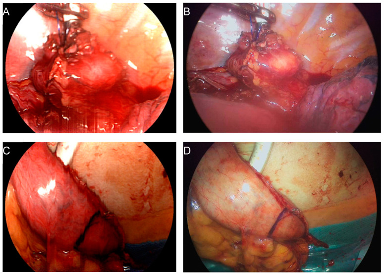
Hyperspectral imaging (HSI) is a non-invasive and contactless technique that enables the real-time acquisition of comprehensive information on tissue within the surgical field. In this pilot study, we investigated whether a new HSI system for minimally-invasive surgery, TIVITA® Mini (HSI-MIS), provides reliable insights into tissue perfusion of the proximal and distal esophagogastric anastomotic sites during 21 laparoscopic/thoracoscopic or robotic Ivor Lewis esophagectomies of patients with cancer to minimize the risk of dreaded anastomotic insufficiency. In this pioneering investigation, physiological tissue parameters were derived from HSI measurements of the proximal site of the anastomosis (esophageal stump) and the distal site of the anastomosis (tip of the gastric conduit) during the thoracic phase of the procedure. Tissue oxygenation (StO2), Near Infrared Perfusion Index (NIR-PI), and Tissue Water Index (TWI) showed similar median values at both anastomotic sites. Significant differences were observed only for NIR-PI (median: 76.5 vs. 63.9; p = 0.012) at the distal site (gastric conduit) compared to our previous study using an HSI system for open surgery. For all 21 patients, reliable and informative measurements were attainable, confirming the feasibility of HSI-MIS to assess anastomotic viability. Further studies on the added benefit of this new technique aiming to reduce anastomotic insufficiency are warranted.

Keywords: clinical evaluation study; esophagectomy; gastrointestinal surgery; hyperspectral imaging; intraoperative imaging; minimally invasive/robotic surgery.


Conflict of interest statement

The authors declare no conflicts of interest.

Figures

Figure 1
Figure 1
Intraoperative imaging with the HSI MIS system. Reconstructed color image based on acquired HSI data (A,C). Corresponding color image provided by the RGB sensor of the same system at video rate (B,D). Imaging of the proximal anastomosis stump after mobilization with inserted stapler head (A,B) and gastric conduit, the distal end of the anastomosis (C,D).
Figure 2
Figure 2
Overview of in vivo studies on HSI of the gastric conduit by our group. (A) Evaluation of open surgery HSI for the measurement of ischemic conditioning effects [28]. (B) Comparison of open surgery HSI and NIR-FI with ICG for perfusion imaging [24]. (C) Validation of the laparoscopic HSI system (HSI MIS) in a controlled setup [32]. (D) Feasibility of intrathoracic HSI and evaluation of the obtained tissue parameters regarding anastomotic sites and HSI systems (this work).
Figure 3
Figure 3
ROI annotation of the distal site of the esophageal anastomosis (gastric conduit). (A) Reconstructed color image from HSI data used to define the ROI (blue line). (B) Corresponding color map of the NIR PI with ROI for statistical analysis.
Figure 4
Figure 4
Motion artifacts due to proximity to the heart. (A) Cardiac motion during the multi-second acquisition process affects the HSI measurement. (B) The corresponding RGB image at video rate is unaffected by cardiac activity.
Figure 5
Figure 5
Distribution of the mean parameter value inside the ROI of the gastroesophageal anastomotic site for all patients obtained by HSI-MIS (Group 1). Circles indicate patients with anastomotic leakage (n = 3). Distal site: tip of the gastric conduit. Proximal site: esophageal remnant stump.
Figure 6
Figure 6
Distribution of the mean parameter value inside the gastric conduit ROI (distal anastomotic site) for all patients measured by HSI-MIS (Group 1) and HSI-Open (Group 2). *: 0.01 < p < 0.05; ns: 0.05 < p (not significant).

Similar articles

Cited by

References

    1. Obermannová R., Alsina M., Cervantes A., Leong T., Lordick F., Nilsson M., van Grieken N.C.T., Vogel A., Smyth E.C. Oesophageal cancer: ESMO Clinical Practice Guideline for diagnosis, treatment and follow-up. Ann. Oncol. Off. J. Eur. Soc. Med. Oncol. 2022;33:992–1004. doi: 10.1016/j.annonc.2022.07.003. - DOI - PubMed
    1. Dehdashti F., Siegel B.A. Neoplasms of the esophagus and stomach. Semin. Nucl. Med. 2004;34:198–208. doi: 10.1053/j.semnuclmed.2004.03.005. - DOI - PubMed
    1. Kato H., Fukuchi M., Miyazaki T., Nakajima M., Tanaka N., Inose T., Kimura H., Faried A., Saito K., Sohda M., et al. Surgical treatment for esophageal cancer. Current issues. Dig. Surg. 2007;24:88–95. doi: 10.1159/000101894. - DOI - PubMed
    1. Kato H., Nakajima M. Treatments for esophageal cancer: A review. Gen. Thorac. Cardiovasc. Surg. 2013;61:330–335. doi: 10.1007/s11748-013-0246-0. - DOI - PubMed
    1. Pu S., Chen H., Zhou C., Yu S., Liao X., Zhu L., He J., Wang B. Major Postoperative Complications in Esophageal Cancer After Minimally Invasive Esophagectomy Compared with Open Esophagectomy: An Updated Meta-analysis. J. Surg. Res. 2021;257:554–571. doi: 10.1016/j.jss.2020.08.011. - DOI - PubMed