Review
. 2024 Jan 7;14(2):136.
doi: 10.3390/diagnostics14020136.

Contemporary Evidence and Practice on Right Heart Catheterization in Patients with Acute or Chronic Heart Failure

Affiliations
Review

Contemporary Evidence and Practice on Right Heart Catheterization in Patients with Acute or Chronic Heart Failure

Lina Manzi et al. Diagnostics (Basel). .

Abstract

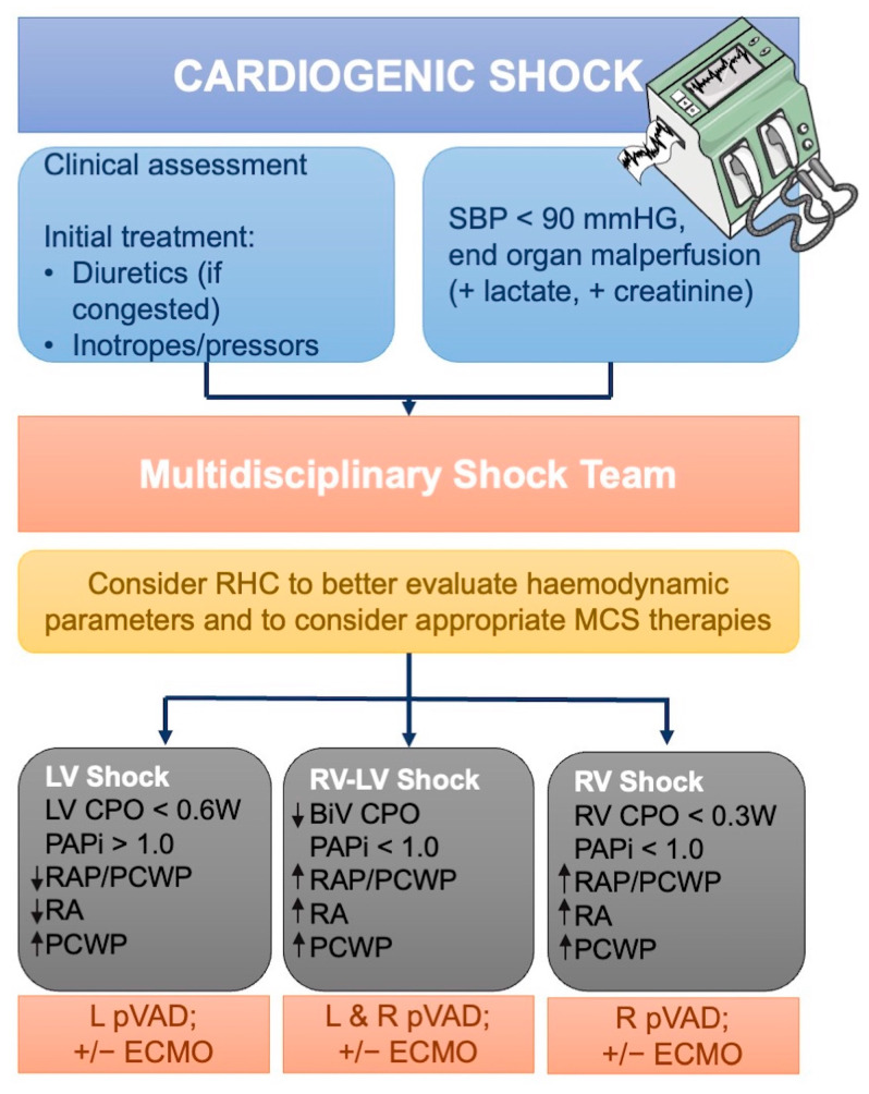
Heart failure (HF) has a global prevalence of 1-2%, and the incidence around the world is growing. The prevalence increases with age, from around 1% for those aged <55 years to >10% for those aged 70 years or over. Based on studies in hospitalized patients, about 50% of patients have heart failure with reduced ejection fraction (HFrEF), and 50% have heart failure with preserved ejection fraction (HFpEF). HF is associated with high morbidity and mortality, and HF-related hospitalizations are common, costly, and impact both quality of life and prognosis. More than 5-10% of patients deteriorate into advanced HF (AdHF) with worse outcomes, up to cardiogenic shock (CS) condition. Right heart catheterization (RHC) is essential to assess hemodynamics in the diagnosis and care of patients with HF. The aim of this article is to review the evidence on RHC in various clinical scenarios of patients with HF.

Keywords: HFpEF; HFrEF; acute heart failure; advanced heart failure; cardiogenic shock; chronic heart failure; heart failure; right heart catheterization.


Conflict of interest statement

The authors declare no conflicts of interest.

Figures

Figure 1
Figure 1
Pressure curves in right heart catheterization. Normal pressure waveforms (a-wave—atrial contraction; c-wave—ventricular contraction; v-wave—atrial filling; x-wave—active atrial emptying; y-wave—passive atrial emptying) obtained in the right atrium (RA), right ventricle (RV), pulmonary artery (PA), and PCWP position with the typical tracings for each; mPAP—mean Pulmonary Artery Pressure; PCWP—Pulmonary Capillary Wedge Pressure; RAP—Right Atrial Pressure; RVDP—Right Ventricular Diastolic Pressure; RVSP—Right Ventricular Systolic Pressure.
Figure 2
Figure 2
HFA-PEFF stepwise diagnostic algorithm. STEP 1 stands for «Pretest assessment»; STEP 2 stands for «Echocardiography and natriuretic peptides score»; STEP 3 stands for «Functional testing in case of uncertainty»; STEP 4 stands for «Final etiology». AF—Atrial Fibrillation; BNP—Brain Natriuretic Peptide; CMR—Cardiac Magnetic Resonance; CO—Cardiac Output; CT—Computed Tomography; ECG—Electrocardiogram; GLS—Global Longitudinal Strain; HF—Heart Failure; HFpEF—Heart Failure with Preserved Ejection Fraction; LAVI—Left Atrial Volume Index; LVMI—Left Ventricular Mass Index; LVWT—Left Ventricular Wall Thickness; NT-proBNP—N-terminal Pro-Brain Natriuretic Peptide; PCWP—Pulmonary Capillary Wedge Pressure; PET—Positron Emission Tomography; RHC—Right Heart Catheterization; RWT—Relative Wall Thickness; TR—Tricuspid Regurgitation; TTE—Transthoracic Echocardiogram; 6MWT—6-Minute Walking Test. Adapted from Pieske B et al. [4].
Figure 3
Figure 3
Representation of pressure invasive exercise RHC. (A). The patient is usually in a supine position, exercising at a cycle ergometer. It is necessary for artery access, usually radial (red arrow), to assess systemic BP and SaO2, and central venous access, usually through a jugular vein (blue arrow), for the procedure itself. A mask (in green) is used to assess ventilatory parameters. (B). Workload scheme. The protocol starts with a cycle unloaded (0 watts) at rest (R) and then starts exercise with feet on a cycle ergometer (F) and increases of 20–25 Watts up to maximal exercise capacity every about 3 min. Measurements are practiced at every stage (X). (C). Representation of mPAP/CO and PCWP/CO slopes. AVO2—Arteriovenous Oxygen Difference; BP—Blood Pressure; CO—Cardiac Output; CO2—Carbon Dioxide; mPAP—Mean Pulmonary Artery Pressure; PA—Pulmonary Artery; PCWP—Pulmonary Capillary Wedge Pressure—SaO2—Oxygen Saturation; RER—Respiratory Exchange Rate; VE—Pulmonary Ventilation during exercise; VCO2—Carbon Dioxide Production; VO2—Venous Oxygen Saturation. Adapted from Hsu S. et al. [10].
Figure 4
Figure 4
Potential role of RHC to support diagnosis and decision-making in the context of advanced heart failure. Diagnostic algorithm of patients with AdHF candidates for heart transplant. With sPAP < 50 mm Hg, PVR < 3 WU, and transpulmonary gradient < 15 mmHg HTX is possible. sPAP ≥ 60 mm Hg, PVR ≥ 5 WU, and transpulmonary gradient ≥ 20 mm Hg are considered prohibitive for transplantation. In patients with 50 mmHg ≤ sPAP < 60 mm Hg, 3 WU ≤ PVR < 5 WU, and 15 mmHg ≤ transpulmonary gradient < 20 mmHg, a vasoreactivity test is indicated. If baseline PVR of ≥3 WU decreases after intravenous sodium nitroprusside to <3 WU, PH is not fixed, and the transplantation is not contraindicated. AdHF—Advanced Heart Failure; HTX—Heart Transplantation; PVR—Pulmonary Vascular Resistance; RHC—Right Heart Catheterization; sPAP—Systolic Pulmonary Artery Pressure; SSAP—Systemic Systolic Arterial Pressure; TPG—Transpulmonary Gradient; VAD—ventricular assist device; VBDT—VAD in Bridge to Decision Therapy; VBTT—VAD in Bridge to Transplantation Therapy; VDT—VAD in Destination Therapy; WU—Wood units. Adapted from Kittleson et al. [86].
Figure 5
Figure 5
Potential role of RHC to support diagnosis and decision-making in the context of cardiogenic shock. A team-based approach to the management of CS. The difference in the type of shock (LV, RV-LV, or RV shock) according to the hemodynamic parameters such as CPO, PAPi, PCWP, CVP, and mean arterial pressure can help the physicians in the choice of the device for ventricular support. BiV—Bi-Ventricular; ECMO—Extracorporeal Membrane Oxygenation; CPO—Cardiac Power Output; LV—Left Ventricular; MCS—Mechanical Circulatory Support; PAPi—Pulmonary Artery Pulsatility Index; PCWP—Pulmonary Capillary Wedge Pressure; pVAD—Percutaneous Ventricular Assist Device; RA—Right Atrial; RAP—Right Atrial Pressure; RHC—Right Heart Catheterization; RV—Right Ventricular; SBP—Systolic Blood Pressure. Adapted from Hsu S. et al. [10].

Similar articles

Cited by

References

    1. McDonagh T.A., Metra M., Adamo M., Gardner R.S., Baumbach A., Böhm M., Burri H., Butler J., Čelutkienė J., Chioncel O., et al. 2021 ESC Guidelines for the Diagnosis and Treatment of Acute and Chronic Heart Failure. Eur. Heart J. 2021;42:3599–3726. doi: 10.1093/eurheartj/ehab368. - DOI - PubMed
    1. Binanay C., Califf R.M., Hasselblad V., O’Connor C.M., Shah M.R., Sopko G., Stevenson L.W., Francis G.S., Leier C.V., Miller L.W., et al. Evaluation Study of Congestive Heart Failure and Pulmonary Artery Catheterization Effectiveness: The ESCAPE Trial. JAMA. 2005;294:1625–1633. doi: 10.1001/jama.294.13.1625. - DOI - PubMed
    1. Kittleson M.M., Panjrath G.S., Amancherla K., Davis L.L., Deswal A., Dixon D.L., Januzzi J.L., Yancy C.W. 2023 ACC Expert Consensus Decision Pathway on Management of Heart Failure with Preserved Ejection Fraction: A Report of the American College of Cardiology Solution Set Oversight Committee. J. Am. Coll. Cardiol. 2023;81:1835–1878. doi: 10.1016/j.jacc.2023.03.393. - DOI - PubMed
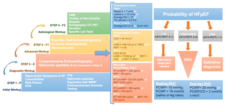
    1. Pieske B., Tschöpe C., de Boer R.A., Fraser A.G., Anker S.D., Donal E., Edelmann F., Fu M., Guazzi M., Lam C.S.P., et al. How to Diagnose Heart Failure with Preserved Ejection Fraction: The HFA-PEFF Diagnostic Algorithm: A Consensus Recommendation from the Heart Failure Association (HFA) of the European Society of Cardiology (ESC) Eur. Heart J. 2019;40:3297–3317. doi: 10.1093/eurheartj/ehz641. - DOI - PubMed
    1. Thakkar A.B., Desai S.P. Swan, Ganz, and Their Catheter: Its Evolution Over the Past Half Century. Ann. Intern. Med. 2018;169:636–642. doi: 10.7326/M17-2145. - DOI - PubMed