Conventional Radiology Evaluation of Neonatal Intravascular Devices (NIVDs): A Case Series
- PMID: 38248034
- PMCID: PMC10814514
- DOI: 10.3390/diagnostics14020157
Conventional Radiology Evaluation of Neonatal Intravascular Devices (NIVDs): A Case Series
Abstract
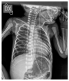
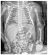
Our radiology department conducted an assessment of 300 neonatal radiographs in the neonatal intensive care unit over almost two years. The purpose was to evaluate the correct positioning of intravascular venous catheters. Our case series revealed that out of a total of 95 cases with misplaced devices, 59 were umbilical venous catheters and 36 were peripherally inserted central catheters. However, all of the central venous catheters were found to be properly positioned. Misplacements of neonatal intravascular devices were found to occur more frequently than expected. The scientific literature contains several articles highlighting the potential complications associated with misplaced devices. Our goal is to highlight the potential misplacements and associated complications that radiologists may encounter while reviewing conventional radiology imaging. Based on our experience, which primarily involved placing UVCs and PICCs, we discovered that conventional radiology is the most effective method for assessing proper device placement with the lowest possible radiation exposure. Given the high number of neonatal vascular device placement procedures, it is essential for radiologists to maintain a high level of vigilance and stay updated on the latest developments in this field.
Keywords: conventional radiology; emergency radiology; neonatal intensive care unit; pediatric emergency radiology; pediatric radiology.
Conflict of interest statement
The authors declare no conflict of interest.
Figures


Similar articles
-
[Radiographic assessment of catheters in a neonatal intensive care unit (NICU)].Rev Chil Pediatr. 2014 Dec;85(6):724-30. doi: 10.4067/S0370-41062014000600011. Rev Chil Pediatr. 2014. PMID: 25697620 Spanish.
-
Sonography for Complete Evaluation of Neonatal Intensive Care Unit Central Support Devices: A Pilot Study.J Ultrasound Med. 2016 Jul;35(7):1465-73. doi: 10.7863/ultra.15.06104. Epub 2016 May 26. J Ultrasound Med. 2016. PMID: 27229130
-
The ABBA project (Assess Better Before Access): A retrospective cohort study of neonatal intravascular device outcomes.Front Pediatr. 2022 Nov 3;10:980725. doi: 10.3389/fped.2022.980725. eCollection 2022. Front Pediatr. 2022. PMID: 36405839 Free PMC article.
-
Focus on peripherally inserted central catheters in critically ill patients.World J Crit Care Med. 2014 Nov 4;3(4):80-94. doi: 10.5492/wjccm.v3.i4.80. eCollection 2014 Nov 4. World J Crit Care Med. 2014. PMID: 25374804 Free PMC article. Review.
-
Intravascular access devices from an interventional radiology perspective: indications, implantation techniques, and optimizing patency.Transfusion. 2018 Feb;58 Suppl 1:549-557. doi: 10.1111/trf.14501. Transfusion. 2018. PMID: 29443411 Review.
References
-
- Alshafei A., Farouk S., Khan A., Ahmed M., Elsaba Y., Aldoky Y. Association of umbilical venous catheters vs peripherally inserted central catheters with death or severe intraventricular hemorrhage among preterm infants < 30 weeks: A randomized clinical trial. J. Neonatal. Perinat. Med. 2023;16:247–255. doi: 10.3233/npm-221126. - DOI - PubMed
LinkOut - more resources
Full Text Sources

