. 2024 Jan 17;14(1):89.
doi: 10.3390/brainsci14010089.

Interleukin-11/IL-11 Receptor Promotes Glioblastoma Cell Proliferation, Epithelial-Mesenchymal Transition, and Invasion

Affiliations

Interleukin-11/IL-11 Receptor Promotes Glioblastoma Cell Proliferation, Epithelial-Mesenchymal Transition, and Invasion

Sarah F Stuart et al. Brain Sci. .

Abstract

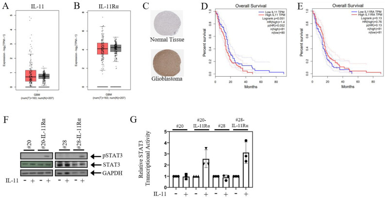
Glioblastoma is highly proliferative and invasive. However, the regulatory cytokine networks that promote glioblastoma cell proliferation and invasion into other areas of the brain are not fully defined. In the present study, we define a critical role for the IL-11/IL-11Rα signalling axis in glioblastoma proliferation, epithelial to mesenchymal transition, and invasion. We identified enhanced IL-11/IL-11Rα expression correlated with reduced overall survival in glioblastoma patients using TCGA datasets. Proteomic analysis of glioblastoma cell lines overexpressing IL-11Rα displayed a proteome that favoured enhanced proliferation and invasion. These cells also displayed greater proliferation and migration, while the knockdown of IL-11Rα reversed these tumourigenic characteristics. In addition, these IL-11Rα overexpressing cells displayed enhanced invasion in transwell invasion assays and in 3D spheroid invasion assays, while knockdown of IL-11Rα resulted in reduced invasion. Furthermore, IL-11Rα-overexpressing cells displayed a more mesenchymal-like phenotype compared to parental cells and expressed greater levels of the mesenchymal marker Vimentin. Overall, our study identified that the IL-11/IL-11Rα pathway promotes glioblastoma cell proliferation, EMT, and invasion.

Keywords: EMT; glioblastoma; interleukin 11; invasion.


Conflict of interest statement

The authors declare no conflicts of interest.

Figures

Figure 1
Figure 1
IL-11Rα signalling correlates with poorer glioblastoma patient survival. Box plots illustrate the expression levels of (A) IL-11 and (B) IL-11Rα in glioblastoma (red) and normal (grey) tissue. (C) Representative immunohistochemistry images of IL-11Rα in glioblastoma and normal brain tissues derived from the HPA database. The relationship between (D) IL-11 and (E) IL-11Rα gene expression and overall patient survival was determined using the TCGA database, with both IL-11 and IL-11Rα gene expression correlated with poorer overall patient survival (p < 0.05). (F) Cells were treated with ±IL-11 for 1 h and then assessed for phospho-STAT3, STAT3, and GAPDH expression by Western blot. (G) Cells were infected with the Ad-APRE-luc adenovirus and, after 24 h, treated with ±IL-11 for a subsequent 24 h. Cells were then lysed and assessed for relative STAT3 transcriptional activity, where untreated cells were normalised to 1.
Figure 2
Figure 2
IL-11Rα expression promotes glioblastoma cell proliferation. (A) #20 and #20-IL-11Rα cells and (B) #28 and #28-IL-11Rα cells were cultured in DME media containing FBS (5%) for three days, then assessed for proliferation using the cell viability assay (n = 3, mean ± SD, where *** indicates p < 0.001 and **** indicates p < 0.0001; Mag = 50×). (C) #20-IL-11Rα and (D) #28-IL-11Rα cells were transfected with control or IL-11Rα siRNA and then cultured in DME media containing FBS (5%) for three days, then assessed for proliferation using the cell viability assay (n = 3, mean ± SD, where ** indicates p < 0.01; Mag = 50×).
Figure 3
Figure 3
IL-11Rα expression promotes glioblastoma cell migration. (A) #20 and #20-IL-11Rα cells and (B) #28 and #28-IL-11Rα cells were cultured in DME media containing FBS (5%) until they formed confluent monolayers. The monolayers were scratched and imaged at 0 and 24 h. Image software analysis was used to determine the percentage of space covered by cells at 24 h (100% = no visible scratch/total wound healed) (n = 3, mean +/− SD, where **** indicates p < 0.0001). (C) #20 and #20-IL-11Rα cells and (D) #28 and #28-IL-11Rα cells were seeded onto a transwell membrane and left for 24 h. The membrane was then fixed and stained with crystal violet (0.25%). Image analysis software was used to determine the percentage of cells stained by the crystal violet after 24 h (n = 3, mean +/− SD, where ** indicates p < 0.01; **** indicates p < 0.001). Representative staining depicts cell migration of cell lines after 24 h (mag = 200×). (E) #20-IL-11Rα and (F) #28-IL-11Rα cells were transfected with control or IL-11Rα siRNA and then seeded onto a transwell membrane and left for 24 h. The membrane was then fixed and stained with crystal violet (0.25%). Image analysis software was used to determine the percentage of cells stained by the crystal violet after 24 h (n = 3, mean +/− SD, where ** indicates p < 0.01; *** indicates p < 0.005). Representative staining depicts cell migration of control and siIL-11Rα transfected cells after 24 h (mag = 200×).
Figure 4
Figure 4
IL-11Rα expression promotes glioblastoma cell invasion. (A) #20 and #20-IL-11Rα and (B) #28 and #28-IL-11Rα cells were seeded onto a transwell membrane coated with Matrigel and left for 24 h. The membrane was then fixed and stained with crystal violet (0.25%). Image analysis software was used to determine the percentage of cells stained by the crystal violet after 24 h (n = 3, mean +/− SD, where *** indicates p < 0.01; **** indicates p < 0.001). Representative staining depicts cell invasion of cell lines after 24 h (mag = 200×). (C) #20-IL-11Rα and (D) #28-IL-11Rα cells were transfected with control or IL-11Rα siRNA and then seeded onto a transwell membrane coated with Matrigel and left for 24 h. The membrane was then fixed and stained with crystal violet (0.25%). Image analysis software was used to determine the percentage of cells stained by the crystal violet after 24 h (n = 3, mean +/− SD, where ** indicates p < 0.01; *** indicates p < 0.005). Representative staining depicts cell invasion of control and siIL-11Rα transfected cells after 24 h (mag = 200×). (E) #20 and #20-IL-11Rα and (F) #28 and #28-IL-11Rα cells were seeded onto non-adherent 24-well plates and left to grow into spheroids. The spheroids were then injected into Matrigel and imaged at 0 and 24 h. Image analysis software was used to determine the relative growth (fold change) of the cell lines over time (n = 3, mean +/− SD, where * indicates p < 0.05; ** indicates p < 0.01). Representative images depict the invasion of #20 and #20-IL-11Rα spheres at 0 and 24 h (mag = 200×).
Figure 5
Figure 5
IL-11Rα expression promotes glioblastoma cell EMT properties. (A) #20, #20-IL-11Rα, #28, and #28-IL-11Rα cells were cultured in DME media containing FBS (5%) and allowed to adhere before being fixed with formalin and stained with crystal violet (0.25%) to observe phenotype (mag = 200×). qPCR was conducted on (B) #20 and #20-IL-11Rα and (C) #28 and #28-IL-11Rα cells to determine the gene expression of Vimentin. The results have been converted to 2ΔΔCT to determine the relative fold change, in which the parental cell line was given a fold change of 1.

Similar articles

Cited by

References

    1. Wang C., Li Y., Chen H., Huang K., Liu X., Qiu M., Liu Y., Yang Y., Yang J. CYP4X1 Inhibition by Flavonoid CH625 Normalizes Glioma Vasculature through Reprogramming TAMs via CB2 and EGFR-STAT3 Axis. J. Pharmacol. Exp. Ther. 2018;365:72–83. doi: 10.1124/jpet.117.247130. - DOI - PubMed
    1. Liu C., Clark P.A., Kuo J.S., Kalejta R.F. Human Cytomegalovirus-Infected Glioblastoma Cells Display Stem Cell-Like Phenotypes. mSphere. 2017;2:e00137-17. doi: 10.1128/mSphere.00137-17. - DOI - PMC - PubMed
    1. Mohme M., Schliffke S., Maire C.L., Runger A., Glau L., Mende K.C., Matschke J., Gehbauer C., Akyuz N., Zapf S., et al. Immunophenotyping of Newly Diagnosed and Recurrent Glioblastoma Defines Distinct Immune Exhaustion Profiles in Peripheral and Tumor-infiltrating Lymphocytes. Clin. Cancer Res. Off. J. Am. Assoc. Cancer Res. 2018;24:4187–4200. doi: 10.1158/1078-0432.CCR-17-2617. - DOI - PubMed
    1. Nakada M., Furuta T., Hayashi Y., Minamoto T., Hamada J. The strategy for enhancing temozolomide against malignant glioma. Front. Oncol. 2012;2:98. doi: 10.3389/fonc.2012.00098. - DOI - PMC - PubMed
    1. McFarland B.C., Hong S.W., Rajbhandari R., Twitty G.B., Jr., Gray G.K., Yu H., Benveniste E.N., Nozell S.E. NF-kappaB-induced IL-6 ensures STAT3 activation and tumor aggressiveness in glioblastoma. PLoS ONE. 2013;8:e78728. doi: 10.1371/journal.pone.0078728. - DOI - PMC - PubMed

LinkOut - more resources