. 2023 Dec 29;46(1):244-261.
doi: 10.3390/cimb46010017.

Characterization of the mIF4G Domains in the RNA Surveillance Protein Upf2p

Affiliations

Characterization of the mIF4G Domains in the RNA Surveillance Protein Upf2p

Edgardo M Colón et al. Curr Issues Mol Biol. .

Abstract

Thirty percent of all mutations causing human disease generate mRNAs with premature termination codons (PTCs). Recognition and degradation of these PTC-containing mRNAs is carried out by the mechanism known as nonsense-mediated mRNA decay (NMD). Upf2 is a scaffold protein known to be a central component of the NMD surveillance pathway. It harbors three middle domains of eukaryotic initiation factor 4G (mIF4G-1, mIF4G-2, mIF4G-3) in its N-terminal region that are potentially important in regulating the surveillance pathway. In this study, we defined regions within the mIF4G-1 and mIF4G-2 that are required for proper function of Upf2p in NMD and translation termination in Saccharomyces cerevisiae. In addition, we narrowed down the activity of these regions to an aspartic acid (D59) in mIF4G-1 that is important for NMD activity and translation termination accuracy. Taken together, these studies suggest that inherently charged residues within mIF4G-1 of Upf2p play a role in the regulation of the NMD surveillance mechanism in S. cerevisiae.

Keywords: Saccharomyces cerevisiae; aspartic acid; cloning; codon; eukaryotic initiation factor-4G; molecular; nonsense; nonsense-mediated mRNA decay.


Conflict of interest statement

The authors declare no conflicts of interest.

Figures

Figure 1
Figure 1
Upf2p was purified by affinity chromatography. (A) Coomassie blue-stained 10% SDS-PAGE of immunopurified Flag-Upf2p which is depicted by an arrow (←). (B) Western blot of immunopurified Flag-Upf2 protein depicted by an arrow (←).
Figure 2
Figure 2
Novel phosphorylation sites were identified in Upf2p mIF4G-1 and mIF4G-2 domains. (A) Example of MS/MS spectrum for precursor ion EPSSSpKETYE. Precursor ion was selected and subjected to fragmentation, generating b and y product ions that represent specific fragments used for identification of the peptide sequence and phosphorylation sites. Loss of H3PO4 from y5 fragment after β-elimination was evidently observed. The double plus sign (++) denotes the charge of the product ion, whereas the asterisk (*) denotes a modification or variation from the usual fragment. (B) Identified phosphorylation sites from the Upf2p precursor ions analyzed by mass spectrometry. First and fourth columns state the starting and ending residues of the precursor ions, respectively. Phosphorylated residues in the second column are depicted by an asterisk (*). Domains containing the peptides are specified in the third column. Last column shows the phosphorylated residue position within Upf2p.
Figure 3
Figure 3
Three phosphorylation sites were identified in Upf2p mIF4G domains. (A) Illustrative representation of Upf2p showing the locations of the MIF4G-1 domain, MIF4G-2 domain, MIF4G-3 domain, acidic domain, and C-Terminal domain. The 12 phosphorylated residues identified by mass spectrometry are presented. (B) S. cerevisiae Upf2p amino acid sequence. Sixty-four percent (64%) of sequence coverage was obtained with the mass spectrometry analysis. Analyzed sequences are shown in gray and phosphorylated residues are in bold.
Figure 4
Figure 4
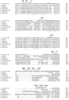
Several residues within Upf2 show a strong conservation of their negative charge. Alignment of sequences from Upf2 S. cerevisiae (NCBI NP_011944.2), S. pombe (NCBI NP_593784.1), A. thaliana (NCBI NP_181459.4), C. elegans (NCBI NP_500974.2), D. melanogaster (NCBI NP_572434.1), H. sapiens (NCBI NP_056357.1), and M. musculus (NCBI NP_001074601.1) shows a conservation of the negative charge of residues D59 and E843 as depicted by the colon (:). Phosphorylated residues are depicted in black while nearby negative residues are in gray. Alignment was constructed with Clustal O (version 1.2.4).
Figure 5
Figure 5
NMD activity requires Upf2p mIF4G-1 and mIF4G-2 phosphorylated regions 1 and 3. (A) Illustrative representation of Upf2p phospho-region mutants (ΔPR). Phosphorylated residues are depicted as black rectangles. In the phospho-region 1 (ΔPR1) residues K50–D59 were deleted, in phospho-region 2 (ΔPR2) residues E321-I330 were deleted, phospho-region 3 (ΔPR3) residues Q421-S430, in phospho-region 4 (ΔPR4) residues G831–Q880 were removed, and in phospho-region 5 (ΔPR5) residues E1019-E1028 were removed. (B) Analysis of cytoplasmic extracts by Western blot showing Upf2p expression. Poly(A) binding protein (Pab1p) was used as a loading control. (C) Northern blot from total cellular RNA was used to determine NMD activity of the Upf2p phospho-region deletions. (D) pre-CYH2 mRNA accumulation from Northern blot expressed as a mean value ± standard deviation. Significance of results, when compared to the WT strain, are represented by asterisks (* p < 0.05, *** p < 0.001).
Figure 6
Figure 6
Proper translation termination requires Upf2p mIF4G-1 and mIF4G-2 phosphorylated regions 1 and 3. can1-100 nonsense suppression assay was used to assess the role of Upf2p in translation termination efficiency. Wild-type and mutant upf2 yeast strains were serially diluted (1:10) five times. Each yeast strain was spotted on SC-Trp-Arg plates, each supplemented with varying concentrations of canavanine (0, 200, or 250 µg/mL), followed by incubation at 30 °C for a duration of two days.
Figure 7
Figure 7
NMD activity does not require Upf2p mIF4G-1 and mIF4G-2 phosphorylated residues of either phospho-region 1 or phospho-region 3. (A) Illustrative representation of Upf2 phospho-region (PR) mutants 1 and 3. Phosphorylated residues S54, S55 and S424 are depicted with an asterisk (*) while adjacent potential phosphorylated residues S52, Y429, S430 are underlined. (B) Analysis of cytoplasmic extracts by Western blot showing Upf2 expression. Poly(A) binding protein (Pab1) was used as a loading control. (C) Northern blot from total cellular RNA was used to determine NMD activity of the Upf2 phosphorylated residues substitutions. (D) pre-CYH2 mRNA accumulation from Northern blot expressed as a mean value ± standard deviation.
Figure 8
Figure 8
Proper translation termination does not require Upf2p mIF4G-1 and mIF4G-2 phosphorylated residues of either phospho-region 1 or phospho-region 3. can1-100 nonsense suppression assay was used to assess the role of Upf2p in translation termination efficiency. Wild-type and mutant upf2 yeast strains were serially diluted (1:10) five times. Each yeast strain was spotted on SC-Trp-Arg plates, each supplemented with varying concentrations of canavanine (0, 200, or 250 µg/ mL), followed by incubation at 30 °C for a duration of two days.
Figure 9
Figure 9
NMD activity requires Upf2p mIF4G-1 and mIF4G-2 non-phosphorylated segments. (A) Illustrative representation of Upf2p non-phosphorylated segments (SR) within phospho-region (PR) 1 and 3. Depicted by a strikethrough, in segment 1 (SR1) residues K50–E53 were deleted, in segment 2 (SR2) residues L57–D59 were deleted, segment 3 (SR3) residues Q42–W423, and in segment 4 (SR4) residues K425–V428 were removed. (B) Analysis of cytoplasmic extracts by Western blot showing Upf2 expression. Poly(A) binding protein (Pab1) was used as a loading control. (C) Northern blot from total cellular RNA was used to determine NMD activity of the Upf2p non-phosphorylated segments deletions. (D) pre-CYH2 mRNA accumulation from Northern blot expressed as a mean value ± standard deviation. Significance of results, when compared to the WT strain, are represented by asterisks (*** p < 0.001).
Figure 10
Figure 10
Proper translation termination requires Upf2p mIF4G-1 and mIF4G-2 non-phosphorylated segments. can1-100 nonsense suppression assay was used to assess the role of Upf2p in translation termination efficiency. Wild-type and mutant upf2 yeast strains were serially diluted (1:10) five times. Each yeast strain was spotted on SC-Trp-Arg plates, each supplemented with varying concentrations of canavanine (0, 200, or 250 µg/mL), followed by incubation at 30 °C for a duration of two days.
Figure 11
Figure 11
NMD activity requires Upf2p mIF4G-1 residue D59. (A) Illustrative representation of Upf2p sub-region mutants. Residues D59, D422, W423, and K425, which were substituted, are depicted with a dagger (†). (B) Analysis of cytoplasmic extracts by Western blot showing Upf2p expression. Poly(A) binding protein (Pab1) was used as a loading control. (C) Northern blot from total cellular RNA was used to determine NMD activity of the Upf2p single mutant deletions. (D) pre-CYH2 mRNA accumulation from Northern blot expressed as a mean value ± standard deviation. Significance of results, when compared to the WT strain, are represented by asterisks (*** p < 0.001).
Figure 12
Figure 12
Proper translation termination requires Upf2p mIF4G-1 residue D59. can1-100 nonsense suppression assay growth curves. Two-way ANOVA was used for statistical analysis. Significance of results, when compared to the WT strain, are represented by asterisks (** p < 0.01, *** p < 0.001).
Figure 13
Figure 13
Characterization of S. cerevisiae Upf2p mIF4G domains. NMD activity requires Upf2p regions and segments within mIF4G-1 and mIF4G-2 and a residue within mIF4G-1. Schematic representation of Upf2p mIF4G domains characterization. mIF4G-1 and mIF4G-2 regions and segments required for NMD are aligned with the numbers (1) and (2), respectively. Single residue within mIF4G-1 required for NMD is aligned with the number (3).

References

    1. Gonzalez C.I., Wang W., Peltz S.W. Nonsense-mediated mRNA decay in Saccharomyces cerevisiae: A quality control mechanism that degrades transcripts harboring premature termination codons. Cold Spring Harb. Symp. Quant. Biol. 2001;66:321–328. doi: 10.1101/sqb.2001.66.321. - DOI - PubMed
    1. Amrani N., Dong S., He F., Ganesan R., Ghosh S., Kervestin S., Li C., Mangus D.A., Spatrick P., Jacobson A. Aberrant termination triggers nonsense-mediated mRNA decay. Biochem. Soc. Trans. 2006;34:39–42. doi: 10.1042/BST0340039. - DOI - PubMed
    1. Behm-Ansmant I., Kashima I., Rehwinkel J., Sauliere J., Wittkopp N., Izaurralde E. mRNA quality control: An ancient machinery recognizes and degrades mRNAs with nonsense codons. FEBS Lett. 2007;581:2845–2853. doi: 10.1016/j.febslet.2007.05.027. - DOI - PubMed
    1. Wang W., Cajigas I.J., Peltz S.W., Wilkinson M.F., Gonzalez C.I. Role for Upf2p phosphorylation in Saccharomyces cerevisiae nonsense-mediated mRNA decay. Mol. Cell Biol. 2006;26:3390–3400. doi: 10.1128/MCB.26.9.3390-3400.2006. - DOI - PMC - PubMed
    1. Kervestin S., Jacobson A. NMD: A multifaceted response to premature translational termination. Nat. Rev. Mol. Cell Biol. 2012;13:700–712. doi: 10.1038/nrm3454. - DOI - PMC - PubMed

LinkOut - more resources