

Significance of Histidine Hydrogen-Deuterium Exchange Mass Spectrometry in Protein Structural Biology
- PMID: 38248468
- PMCID: PMC10813008
- DOI: 10.3390/biology13010037
Significance of Histidine Hydrogen-Deuterium Exchange Mass Spectrometry in Protein Structural Biology
Abstract
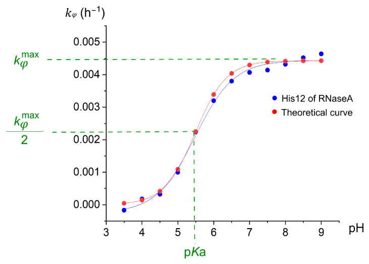
Histidine residues play crucial roles in shaping the function and structure of proteins due to their unique ability to act as both acids and bases. In other words, they can serve as proton donors and acceptors at physiological pH. This exceptional property is attributed to the side-chain imidazole ring of histidine residues. Consequently, determining the acid-base dissociation constant (Ka) of histidine imidazole rings in proteins often yields valuable insights into protein functions. Significant efforts have been dedicated to measuring the pKa values of histidine residues in various proteins, with nuclear magnetic resonance (NMR) spectroscopy being the most commonly used technique. However, NMR-based methods encounter challenges in assigning signals to individual imidazole rings and require a substantial amount of proteins. To address these issues associated with NMR-based approaches, a mass-spectrometry-based method known as histidine hydrogen-deuterium exchange mass spectrometry (His-HDX-MS) has been developed. This technique not only determines the pKa values of histidine imidazole groups but also quantifies their solvent accessibility. His-HDX-MS has proven effective across diverse proteins, showcasing its utility. This review aims to clarify the fundamental principles of His-HDX-MS, detail the experimental workflow, explain data analysis procedures and provide guidance for interpreting the obtained results.
Keywords: acid dissociation constant; histidine; histidine protonation; mass spectrometry; protein structural biology.
Conflict of interest statement
The authors declare no conflicts of interest.
Figures


Similar articles
-
Histidine hydrogen-deuterium exchange mass spectrometry for probing the microenvironment of histidine residues in dihydrofolate reductase.PLoS One. 2011 Feb 16;6(2):e17055. doi: 10.1371/journal.pone.0017055. PLoS One. 2011. PMID: 21359214 Free PMC article.
-
Analysis of conformational changes in rhodopsin by histidine hydrogen-deuterium exchange.Methods Mol Biol. 2015;1271:123-32. doi: 10.1007/978-1-4939-2330-4_9. Methods Mol Biol. 2015. PMID: 25697521
-
Determination of pKa values of individual histidine residues in proteins using mass spectrometry.Anal Chem. 2008 Sep 1;80(17):6481-7. doi: 10.1021/ac8009643. Epub 2008 Jul 30. Anal Chem. 2008. PMID: 18665614
-
Measuring the hydrogen/deuterium exchange of proteins at high spatial resolution by mass spectrometry: overcoming gas-phase hydrogen/deuterium scrambling.Acc Chem Res. 2014 Oct 21;47(10):3018-27. doi: 10.1021/ar500194w. Epub 2014 Aug 29. Acc Chem Res. 2014. PMID: 25171396 Review.
-
Computational Structure Prediction for Antibody-Antigen Complexes From Hydrogen-Deuterium Exchange Mass Spectrometry: Challenges and Outlook.Front Immunol. 2022 May 26;13:859964. doi: 10.3389/fimmu.2022.859964. eCollection 2022. Front Immunol. 2022. PMID: 35720345 Free PMC article. Review.
Cited by
-
Deuterium trafficking, mitochondrial dysfunction, copper homeostasis, and neurodegenerative disease.Front Mol Biosci. 2025 Jul 22;12:1639327. doi: 10.3389/fmolb.2025.1639327. eCollection 2025. Front Mol Biosci. 2025. PMID: 40765848 Free PMC article. Review.
-
Hydrogen/Deuterium Exchange and Protein Oxidative Footprinting with Mass Spectrometry Collectively Discriminate the Binding of Small-Molecule Therapeutics to Bcl-2.Anal Chem. 2025 Mar 4;97(8):4329-4340. doi: 10.1021/acs.analchem.4c04516. Epub 2025 Feb 19. Anal Chem. 2025. PMID: 39969248
-
Is Deuterium Sequestering by Reactive Carbon Atoms an Important Mechanism to Reduce Deuterium Content in Biological Water?FASEB Bioadv. 2025 May 14;7(6):e70019. doi: 10.1096/fba.2025-00032. eCollection 2025 Jun. FASEB Bioadv. 2025. PMID: 40496345 Free PMC article. Review.
References
-
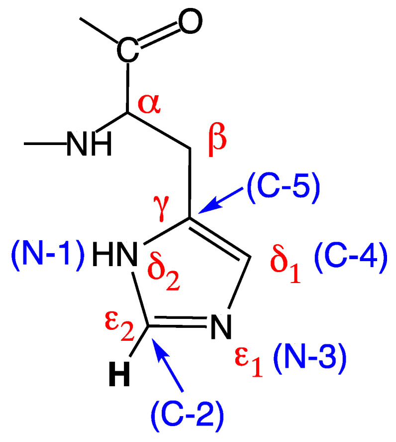
- Nomenclature and Symbolism for Amino Acids and Peptides (Recommendations 1983) Pure Appl. Chem. 1984;56:595–624. doi: 10.1351/pac198456050595. - DOI
-
- Markley J.L. Observation of Histidine Residues in Proteins by Nuclear Magnetic Resonance Spectroscopy. Acc. Chem. Res. 1975;8:70–80. doi: 10.1021/ar50086a004. - DOI
Publication types
LinkOut - more resources
Full Text Sources
