Review
. 2023 Dec 23;14(1):12.
doi: 10.3390/metabo14010012.

Lipid Metabolism in Metabolic-Associated Steatotic Liver Disease (MASLD)

Affiliations
Review

Lipid Metabolism in Metabolic-Associated Steatotic Liver Disease (MASLD)

Majid Mufaqam Syed-Abdul. Metabolites. .

Abstract

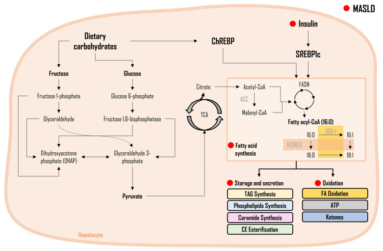
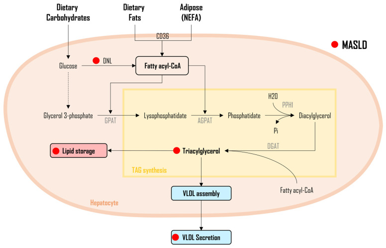
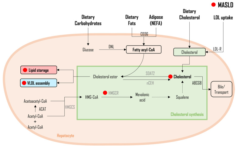
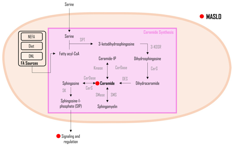
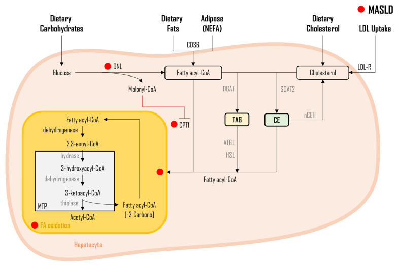
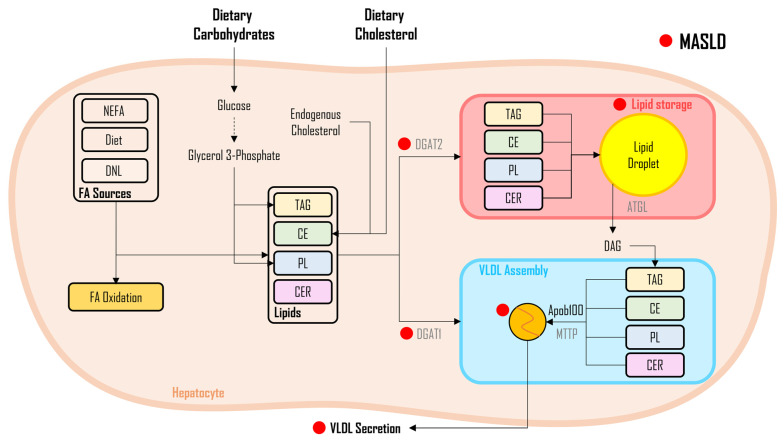
Metabolic-associated steatotic liver disease (MASLD) is a cluster of pathological conditions primarily developed due to the accumulation of ectopic fat in the hepatocytes. During the severe form of the disease, i.e., metabolic-associated steatohepatitis (MASH), accumulated lipids promote lipotoxicity, resulting in cellular inflammation, oxidative stress, and hepatocellular ballooning. If left untreated, the advanced form of the disease progresses to fibrosis of the tissue, resulting in irreversible hepatic cirrhosis or the development of hepatocellular carcinoma. Although numerous mechanisms have been identified as significant contributors to the development and advancement of MASLD, altered lipid metabolism continues to stand out as a major factor contributing to the disease. This paper briefly discusses the dysregulation in lipid metabolism during various stages of MASLD.

Keywords: DNL; MASH; MASLD; cholesterol; fatty acid oxidation.


Conflict of interest statement

The authors declare no conflict of interest.

Figures

Figure 1
Figure 1
Fatty acid synthesis and regulation in the development of MASLD. Legend: This figure illustrates the intricate process of Fatty Acid (FA) synthesis and its regulation. The synthesis of FAs involves a complex biochemical pathway, primarily driven by the activities of key enzymes such as ACC; FASN; SCD-1; and elongases (ELOV). The master regulator SREBP1c, influenced by insulin levels, governs the activation of enzymes in de novo lipogenesis (DNL). Additionally, ChREBP serves as a regulator activated during postprandial states and hyperglycemia, impacting gene transcription and enzymatic activity in the DNL pathway. The red dot (●) represents changes altered during MASLD pathogenesis. Figure abbreviations: ACC—acetyl-CoA carboxylase; CE—cholesterol ester; ELOVL5—elongases 5; FASN—fatty acid synthase; SCD-1—stearoyl-CoA desaturase-1; TCA—tricarboxylic cycle; and TAG—triacylglycerols.
Figure 2
Figure 2
Triacylglycerol and diacylglycerol synthesis in MASLD. Legend: This figure illustrates the general metabolism of triacylglycerol (TAG) and diacylglycerol (DAG) synthesis. Three distinct sources of fatty acids (FAs) undergo enzymatic conversions, leading to the formation of fatty acyl-CoA. Notably, in MASLD, elevated DAG levels contribute to lipotoxicity, exacerbating fat accumulation, oxidative stress, inflammation, and insulin resistance. Mechanistically, DAG inhibits insulin receptor tyrosine kinase via PKC-ε activation, underscoring its significance in MASLD pathogenesis. Increased triacylglycerol (TAG) levels signify increased hepatic TAG accumulation, marking a crucial stage in MASLD progression. The red dot (●) represents changes altered during MASLD pathogenesis. Figure abbreviations: AGPAT—1-acylglycerol-3-phosphate-O-acyltransferase; CD36—cluster of differentiation 36; CE—cholesterol ester; DGAT—diacylglycerol acyltransferase enzyme; DNL—de novo lipogenesis; FA—fatty acids; GPAT—glycerol-3-phosphate; Pi—phosphate; NEFA—nonesterified fatty acids; PDAT—Phospholipid: diacylglycerol acyltransferase; PPH-1—phosphohydrolase–1; TAG—triacylglycerols; and VLDL—very-low-density lipoprotein.
Figure 3
Figure 3
Cholesterol synthesis and implications in MASLD. Legend: This figure outlines cholesterol synthesis, with acetyl-CoA acetyltransferases initiating the pathway and HMG-CoA reductase progressing to cholesterol. In MASLD, elevated total cholesterol and LDLc, along with reduced HDLc, are observed. SREBP-2, a master regulator, shows increased expression, contributing to heightened cholesterol synthesis. Enhanced HMGCR activity in MASLD and MASH exacerbates cholesterol production, impacting liver function. Altered cholesterol metabolism plays a crucial role in MASLD progression, with potential implications for inflammation and fibrosis within liver cells. The red dot (●) represents changes altered during MASLD pathogenesis. Figure abbreviations: ABCG8—ATP-binding cassette sub-family G member 8; ACAT—acetyl-CoA acetyltransferases; DNL—de novo lipogenesis; HMGCR—β-Hydroxy β-methylglutaryl-CoA reductase; HMGCS—β-Hydroxy β-methylglutaryl-CoA synthase; LDL-R—low-density lipoprotein receptor; NCEH1—neutral cholesterol ester hydrolase 1; NEFA—nonesterified fatty acids; and SOAT2—sterol O-acyltransferase 2.
Figure 4
Figure 4
Ceramide metabolism in MASLD. Legend: This figure outlines ceramide synthesis and impacts, emphasizing crucial roles in cell membrane integrity, skin barrier function, apoptosis, and lipid metabolism. Elevated ceramide levels in metabolic disorders, cardiovascular diseases, and MASLD are linked to increased lipid peroxidation and reduced mitochondrial respiration in MASH. Ceramides contribute significantly to hepatic steatosis and insulin resistance by activating PKCζ and PP2A, impairing AKT translocation, suppressing β-oxidation, and activating the NLRP3 inflammasome. The red dot (●) represents changes altered during MASLD pathogenesis. Figure abbreviations: 3-KDSR—3-ketodihydrosphinganine reductase; C1P—ceramide-1-phosphate; Cer—ceramides; CerDase—ceramidase; CerS—ceramide synthase; DES—dihydroceramide desaturase; S1P—sphingosine-1-phosphate; SK—sphingosine kinase; SMase—sphingomyelinase; SMS—sphingomyelin synthase; and SPT—serine palmitoyltransferase.
Figure 5
Figure 5
Fatty acid oxidation in MASLD. Legend: Illustration of diverse fatty acid sources contributing to cellular oxidation in non-alcoholic fatty liver disease (MASLD). Carnitine palmitoyltransferase 1 (CPT1), a pivotal outer-membrane mitochondrial enzyme, facilitates fatty acid uptake and oxidation through the ß-oxidation pathway. Malonyl-CoA, an intermediate in de novo lipogenesis (DNL), inhibits CPT1, preventing the entry of fatty acids from plasma non-esterified fatty acids (NEFA), diet, triglyceride (TAG) catabolism, and cholesterol ester (CE) hydrolysis into mitochondria for oxidation. This dysregulation results in hepatic lipid accumulation, fostering the progression of liver steatosis and metabolic dysfunction. The red dot (●) represents changes altered during MASLD pathogenesis. Figure abbreviations: 2C—two carbons; ATGL—Adipose triglyceride lipase; CD36—cluster of differentiation 36; CE—cholesterol esters; CPT1—carnitine palmitoyltransferase 1; DGAT—diacylglycerol acyltransferase enzyme; DNL—de novo lipogenesis; FA—fatty acids; HSL—hormone-sensitive lipase; LDL-R—low-density lipoprotein receptor; MTP—mitochondrial trifunctional protein; NCEH—neutral cholesterol ester hydrolase; NEFA—non-esterified fatty acids; SOAT2—sterol O-acyltransferase 2; and TAG—triacylglycerols.
Figure 6
Figure 6
Lipid flux in MASLD. Legend: This figure illustrates the intricate fate of fatty acids (FAs). FAs from de novo lipogenesis (DNL), non-esterified FAs (NEFA), and dietary fats undergo oxidation for energy or are utilized in lipid biosynthesis (triglycerides [TAG], ceramides [CER], and phospholipids [PL]). While DGAT1 activation drives FA incorporation into lipoprotein particles (VLDL) for circulation, DGAT2 activation leads to storage in hepatic lipid droplets. Insulin resistance in MASLD diminishes insulin-induced suppression of VLDL synthesis, contributing to elevated VLDL secretion and subsequent lipid dysregulation. The red dot (●) represents changes altered during MASLD pathogenesis. Figure abbreviations: APOB100—apolipoprotein B100; ATGL—adipose triglyceride lipase; CE—cholesterol esters; DAG—diacylglycerols; DGAT—diacylglycerol acyltransferase enzyme; DNL—de novo lipogenesis; FA—fatty acids; MTTP—microsomal triglyceride transfer protein; NEFA—nonesterified fatty acids; PL—phospholipids; TAG—triacylglycerols; and VLDL—very-low-density lipoprotein.

Similar articles

Cited by

References

    1. Rinella M.E., Lazarus J.V., Ratziu V., Francque S.M., Sanyal A.J., Kanwal F., Romero D., Abdelmalek M.F., Anstee Q.M., Arab J.P., et al. A multisociety Delphi consensus statement on new fatty liver disease nomenclature. Hepatology. 2023;78:1966–1986. doi: 10.1097/HEP.0000000000000520. - DOI - PMC - PubMed
    1. Sanyal A.J., American Gastroenterological A. AGA technical review on nonalcoholic fatty liver disease. Gastroenterology. 2002;123:1705–1725. doi: 10.1053/gast.2002.36572. - DOI - PubMed
    1. Younossi Z., Anstee Q.M., Marietti M., Hardy T., Henry L., Eslam M., George J., Bugianesi E. Global burden of NAFLD and NASH: Trends, predictions, risk factors and prevention. Nat. Rev. Gastroenterol. Hepatol. 2018;15:11–20. doi: 10.1038/nrgastro.2017.109. - DOI - PubMed
    1. Younossi Z.M., Koenig A.B., Abdelatif D., Fazel Y., Henry L., Wymer M. Global epidemiology of nonalcoholic fatty liver disease-Meta-analytic assessment of prevalence, incidence, and outcomes. Hepatology. 2016;64:73–84. doi: 10.1002/hep.28431. - DOI - PubMed
    1. Fazel Y., Koenig A.B., Sayiner M., Goodman Z.D., Younossi Z.M. Epidemiology and natural history of non-alcoholic fatty liver disease. Metabolism. 2016;65:1017–1025. doi: 10.1016/j.metabol.2016.01.012. - DOI - PubMed

LinkOut - more resources