

Electrocardiographic Changes with Age in Japanese Patients with Noonan Syndrome
- PMID: 38248880
- PMCID: PMC10816141
- DOI: 10.3390/jcdd11010010
Electrocardiographic Changes with Age in Japanese Patients with Noonan Syndrome
Abstract
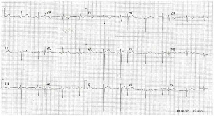
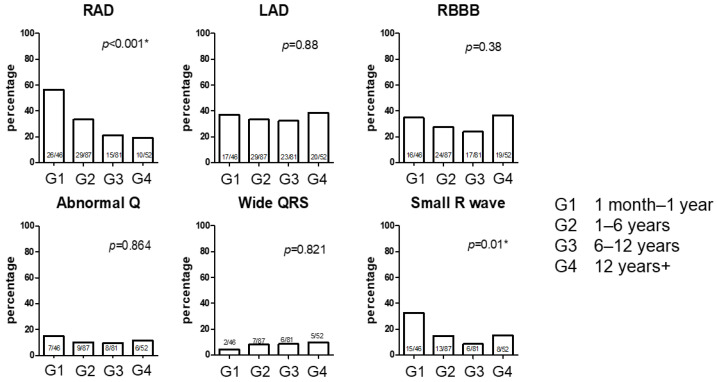
Little information is available on age-related electrocardiographic changes in patients with Noonan syndrome. This single-center study evaluated the electrocardiograms of patients with Noonan syndrome. We divided the patients (n = 112; electrocardiograms, 256) into four groups according to age: G1 (1 month-1 year), G2 (1-6 years), G3 (6-12 years), and G4 (>12 years). Typical Noonan syndrome-related electrocardiographic features such as left-axis deviation, abnormal Q wave, wide QRS complex, and small R wave in precordial leads were detected. A high percentage of QRS axis abnormalities was found in all groups. Significant differences in right-axis deviation (RAD) were noted among the groups: 56.5% of G1 patients showed RAD compared with 33.3% of G2, 21.1% of G3, and 19.2% of G4 patients. The small R was also significantly different among the groups: 32.6% of G1 patients showed a small R wave compared with 14.9% of G2, 8.5% of G3, and 15.4% of G4 patients. Of the 53 patients with Noonan syndrome aged 1 month to 2 years, 18 had T-positive V1 with a higher prevalence of pulmonary stenosis and cardiac interventions. QRS axis abnormalities, small R in V6, and T-positive V1 could help diagnose Noonan syndrome in infants or young children.
Keywords: Noonan syndrome; T-positive V1; electrocardiogram; right-axis deviation; small R.
Conflict of interest statement
The authors declare no conflicts of interest.
Figures
Similar articles
-
The added value of the electrocardiogram in Noonan syndrome.Cardiol Young. 2022 Jun;32(6):936-943. doi: 10.1017/S1047951121003310. Epub 2021 Aug 12. Cardiol Young. 2022. PMID: 34382536
-
Differentiating primary sarcomeric hypertrophic cardiomyopathy from Noonan syndrome: can the electrocardiogram be of use?Cardiol Young. 2024 Mar;34(3):597-603. doi: 10.1017/S1047951123003177. Epub 2023 Aug 31. Cardiol Young. 2024. PMID: 37649442
-
Electrocardiography in Noonan syndrome PTPN11 gene mutation--phenotype characterization.Am J Med Genet A. 2008 Feb 1;146A(3):350-3. doi: 10.1002/ajmg.a.32140. Am J Med Genet A. 2008. PMID: 18203203
-
[Wide QRS complex tachycardia: an old and new problem].G Ital Cardiol (Rome). 2009 Sep;10(9):580-95. G Ital Cardiol (Rome). 2009. PMID: 19891250 Review. Italian.
-
Usefulness of the 12-lead electrocardiogram in the follow-up of patients with cardiac resynchronization devices. Part I.Cardiol J. 2011;18(5):476-86. doi: 10.5603/cj.2011.0002. Cardiol J. 2011. PMID: 21947982 Review.
Cited by
-
The NRF2/ID2 Axis in Vascular Smooth Muscle Cells: Novel Insights into the Interplay between Vascular Calcification and Aging.Aging Dis. 2024 May 20;16(2):1120-1140. doi: 10.14336/AD.2024.0075. Aging Dis. 2024. PMID: 38916733 Free PMC article.
-
X-Chromosome-Linked miRNAs Regulate Sex Differences in Cardiac Physiology.Circ Res. 2025 Jan 31;136(3):258-275. doi: 10.1161/CIRCRESAHA.124.325447. Epub 2024 Dec 30. Circ Res. 2025. PMID: 39772608
-
Broad Electrocardiogram Syndromes Spectrum: From Common Emergencies to Particular Electrical Heart Disorders-Part II.Diagnostics (Basel). 2025 Jun 19;15(12):1568. doi: 10.3390/diagnostics15121568. Diagnostics (Basel). 2025. PMID: 40564888 Free PMC article. Review.
References
-
- Baban A., Olivini N., Lepri F.R., Calì F., Mucciolo M., Digilio M.C., Calcagni G., di Mambro C., Dallapiccola B., Adorisio R., et al. SOS1 mutations in Noonan syndrome: Cardiomyopathies and not only congenital heart defects! Report of six patients including two novel variants and literature review. Am. J. Med. Genet. A. 2019;179:2083–2090. doi: 10.1002/ajmg.a.61312. - DOI - PubMed
Grants and funding
LinkOut - more resources
Full Text Sources
