. 2023 Dec 29;10(1):25.
doi: 10.3390/jof10010025.

Identification and Characterization of a Predominant Hydrophobin in the Edible Mushroom Grifola frondosa

Affiliations

Identification and Characterization of a Predominant Hydrophobin in the Edible Mushroom Grifola frondosa

Bo Song et al. J Fungi (Basel). .

Abstract

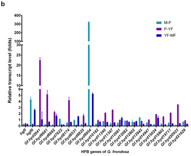
Hydrophobins (HFBs) are a group of small, secreted amphipathic proteins of fungi with multiple physiological functions and potential commercial applications. In this study, HFB genes of the edible mushroom, Grifola frondosa, were systematically identified and characterized, and their transcriptional profiles during fungal development were determined. In total, 19 typical class I HFB genes were discovered and bioinformatically analyzed. Gene expression profile examination showed that Gf.hyd9954 was particularly highly upregulated during primordia formation, suggesting its major role as the predominant HFB in the lifecycle of G. frondosa. The wettability alteration profile and the surface modification ability of recombinant rGf.hyd9954 were greater than for the Grifola HFB HGFII-his. rGf.hyd9954 was also demonstrated to form the typical class I HFB characteristic-rodlet bundles. In addition, rGf.hyd9954 was shown to possess nanoparticle characteristics and emulsification activities. This research sheds light on the regulation of fungal development and its association with the expression of HFB genes.

Keywords: fungal development; hydrophobins; life cycle; maitake; transcription levels.


Conflict of interest statement

The authors declare that they have no competing interests.

Figures

Figure 1
Figure 1
The primary gene structure and phylogeny analysis of hydrophobin genes among G. frondosa. (a) Gene structure of the hydrophobin genes. PF01185 is the domain of “fungal hydrophobin”. (b) Phylogeny between two strains and their putative hydrophobin genes. G. frondosa CICC®50075 and 9006-11 are the two sub-lineages of G. frondosa. In order to analyze the hydrophobin genes of these two strains and their own phylogeny, the Neighbor-Joining Tree (1000 bootstrap replicates) was conducted via MEGA7 software. Putative hydrophobin genes Gf.hydXXXX are from CICC®50075, while the others are from lineage 9006-11.
Figure 2
Figure 2
The expression pattern of hydrophobin genes during one-life cycle of G. frondosa CICC®50075. (a) The relative expression levels of typical hydrophobin genes at the mycelia, primordial stage, and fruiting body development stages; all the expression levels were calibrated with the levels of the mycelia stage. S1, the mycelia (Myc) stage; S2, the primordial stage (Pri); S3, the young fruiting body (Yfb) stage; S4 represents the mature fruiting body (Mfb) stage. (b) The relative expression levels of hydrophobin genes that compared to former stage. * represents p < 0.05, ** represents p < 0.01, ns represents nonsignificant difference.
Figure 2
Figure 2
The expression pattern of hydrophobin genes during one-life cycle of G. frondosa CICC®50075. (a) The relative expression levels of typical hydrophobin genes at the mycelia, primordial stage, and fruiting body development stages; all the expression levels were calibrated with the levels of the mycelia stage. S1, the mycelia (Myc) stage; S2, the primordial stage (Pri); S3, the young fruiting body (Yfb) stage; S4 represents the mature fruiting body (Mfb) stage. (b) The relative expression levels of hydrophobin genes that compared to former stage. * represents p < 0.05, ** represents p < 0.01, ns represents nonsignificant difference.
Figure 3
Figure 3
General XPS spectrum of blank silicon slice surfaces and hydrophobin-containing silicon slice surfaces. Hydrophobin-containing silicon slice surfaces XPS spectra of rGf.hyd9954 and HGFII-his.
Figure 4
Figure 4
The characterization of the rGf.hyd9954 assembly. (a) The ThT analysis of the rGf.hyd9954. ThT is a fluorescent dye that is commonly used for monitoring the amyloid formation by proteins and peptides. The hydrophobin assembly frequently accompanied by the formation of the cross-β structure. The emission intensity of ThT dye at 480 nm (excitation at 450 nm) increases significantly when it binds to the cross β structure of hydrophobins. (b) The morphology detection of the rGf.hyd9954 assembly with AFM. The surface topography of rGf.hyd9954-modified mica was carried out. From the image, we confirmed the rGf.hyd9954, like any class I hydrophobins, is able to self-assemble into amyloid-like fibrils with lengths in the range of 100–150 nm.
Figure 5
Figure 5
The hydropathy plot analysis, surface hydrophobicity measurements and the putative tertiary structure. (a) The hydropathy plot analysis was performed by the Kyte and Doolittle method to analyze the hydrophobicity of proteins. (b) The fluorescence spectrum of 1,8-ANS in the presence of rGf.hyd9954 and the spectrum of HGFII-his. (c) The automated protein structure homology modeling of rGf.hyd9954 on Swiss-model (https://swissmodel.expasy.org/, accessed on 18 September 2023). Homology-based three-dimensional model of the solution NMR structure of a class I hydrophobin from Serpula lacrymans (5w0y.1.A).
Figure 6
Figure 6
The particle size assay. The particle sizes were carried out by the dynamic light scattering (DLS) method to analyze the particle size of rGf.hyd9954 solutions at 100 μg/mL.
Figure 7
Figure 7
The emulsification properties determination of rGf,hyd9954. (a) The ddH2O-soybean oil; (b) the BSA solution-soybean oil; (c) the rGf.hyd9954-soybean oil; and (d) the HGFII-his-soybean oil emulsification systems. The protein concentration of the protein solutions was 100 μg/mL. The insertion figures were the emulsification systems contained in a transparent vial after the homogenization treatment for 72 h, bar 5 μm.

Similar articles

Cited by

References

    1. Schor M., Reid J.L., MacPhee C.E., Stanley-Wall N.R. The Diverse Structures and Functions of Surfactant Proteins. Trends Biochem. Sci. 2016;41:610–620. doi: 10.1016/j.tibs.2016.04.009. - DOI - PMC - PubMed
    1. De Vries O.M., Moore S., Arntz C., Wessels J.G., Tudzynski P. Identification and characterization of a tri-partite hydrophobin from Claviceps fusiformis. A novel type of class II hydrophobin. Eur. J. Biochem. 1999;262:377–385. doi: 10.1046/j.1432-1327.1999.00387.x. - DOI - PubMed
    1. Wessels J.G.H. Fungal hydrophobins: Proteins that function at an interface. Trends Plant Sci. 1996;1:9–15. doi: 10.1016/S1360-1385(96)80017-3. - DOI
    1. Wösten H.A., de Vocht M.L. Hydrophobins, the fungal coat unravelled. Biochim. Biophys. Acta. 2000;1469:79–86. doi: 10.1016/S0304-4157(00)00002-2. - DOI - PubMed
    1. Shin Y.K., Kim D.W., Lee S.W., Lee M.J., Gi Baek S., Lee T., Yun S.H. Functional roles of all five putative hydrophobin genes in growth, development, and secondary metabolism in Fusarium graminearum. Fungal Genet. Biol. 2022;160:103683. doi: 10.1016/j.fgb.2022.103683. - DOI - PubMed

LinkOut - more resources