. 2024 Jan 7;10(1):54.
doi: 10.3390/jof10010054.

Hyperpolarisation of Mitochondrial Membranes Is a Critical Component of the Antifungal Mechanism of the Plant Defensin, Ppdef1

Affiliations

Hyperpolarisation of Mitochondrial Membranes Is a Critical Component of the Antifungal Mechanism of the Plant Defensin, Ppdef1

Kathy Parisi et al. J Fungi (Basel). .

Abstract

Plant defensins are a large family of small cationic proteins with diverse functions and mechanisms of action, most of which assert antifungal activity against a broad spectrum of fungi. The partial mechanism of action has been resolved for a small number of members of plant defensins, and studies have revealed that many act by more than one mechanism. The plant defensin Ppdef1 has a unique sequence and long loop 5 with fungicidal activity against a range of human fungal pathogens, but little is known about its mechanism of action. We screened the S. cerevisiae non-essential gene deletion library and identified the involvement of the mitochondria in the mechanism of action of Ppdef1. Further analysis revealed that the hyperpolarisation of the mitochondrial membrane potential (MMP) activates ROS production, vacuolar fusion and cell death and is an important step in the mechanism of action of Ppdef1, and it is likely that a similar mechanism acts in Trichophyton rubrum.

Keywords: Ppdef1; S. cerevisiae; Trichophyton rubrum; antifungal; hyperpolarisation; plant defensin.


Conflict of interest statement

N.L.v.d.W., J.A.M. and M.A.A. are Hexima Ltd. shareholders. N.L.v.d.W. and M.A.A. are inventors of the method for the treatment of fungal infections, EP3209319B1 and US9713632B2. Hexima Ltd. has granted permission to publish the results. We confirm that neither the manuscript nor any parts of its content are currently under consideration or published in another journal. Authors K.P., J.A.M., R.L., K.S.H., R.G., E.L., N.L.v.d.W., M.R.B. and M.A.A. were employed by the company Hexima Ltd. The remaining authors declare that the research was conducted in the absence of any commercial or financial relationships that could be construed as a potential conflict of interest.

Figures

Figure 1
Figure 1
Ppdef1 shares little sequence similarity with other plant defensins. (A) Percent conservation of residues between the ~1000 plant defensin sequences. The black lines connecting the conserved cysteine residues represent the disulfide bonds. Cysteine residues are highlighted in deep blue. (B) MUSCLE alignment of native sequences for Ppdef1 to plant defensins with partially characterised mechanisms of action. Conserved residues are coloured deep purple, and similar amino acids are light purple. Gaps have been inserted to maximize alignment. Sequence identity and predicted charge of the loop 5 sequence at pH 7.0 are listed on the right. ‘.’ Indicates residues with weakly similar properties, and ‘:’ indicates residues with strongly similar properties. The black line beneath the alignment shows regions between cysteine residues defined as loops 1–7 (Ppdef1—Picramnia pentandra; NaD1—Nicotiana alata Q8GTM0; ZmD32—Triticum aestivum 6DMZ_A; NbD6—Nicotiana benthamiana [16]; NaD2—Nicotiana alata AOD75394; DmAMP1—Dahlia merkii AAB34972; RsAFP2—Raphanus sativus P30230; MtDef4—Medicago truncatula 2LR3_A; and HsAFP1—Heuchera sanguinea AAB34974.1).
Figure 2
Figure 2
Lipid strip and carbohydrate binding. (A) PIP stripTM probed with NaD1 and Ppdef1. NaD1 bound to PI(3,4)P2, PI(4,5)P2, PI(3,4,5)P3, PA and PI(5)P, and Ppdef1 did not bind. NaD1 and Ppdef1 (0.025 μg) were used as positive controls. (B) SphingoStripTM probed with DmAMP1 and Ppdef1. DmAMP1 and Ppdef1 did not bind to any of the lipids on the SphingoStripTM. Purified defensins (0.025 μg) were used as positive controls. (C,D) Amount of defensin remaining in the supernatant after incubation with increasing amounts of insoluble polysaccharide. Defensins were detected using Western blot with specific antibodies. (C) NaD1, NaD2, DmAMP1 and Ppdef1 binding to chitin. (D) NaD1, Ppdef1, NaD2 and DmAMP1 binding to yeast β-glucan.
Figure 3
Figure 3
Uptake of Ppdef1-FAM into S. cerevisiae cells, effect on ROS production, vacuole morphology and cell permeability. S. cerevisiae cells, pre-labelled with FM-464 vacuolar stain (red), were treated with Ppdef1-FAM (green, (A,B,E,F)) or unlabelled Ppdef1 (C,D) in a CellASIC ONIX microfluidic plate. The labelled defensin (green), vacuolar morphology (red), ROS (green) and permeabilization (blue) were monitored over time using confocal microscopy. In one experiment, the uptake of Ppdef1-FAM was monitored (A,B). Ppdef1-FAM bound to the surface of the cell ((Ai) red and green merged channels) within 11 min and had moved into the cytoplasm ((Aii) green channel only) highlighted with yellow arrows. At ~16 min, the vacuoles had fused into one large vacuole ((Bi) red and green merged channels; (Bii) red channel only) from multiple smaller vacuoles ((Ai) red and green merged channels) highlighted with green arrows. As the vacuoles fused, Ppdef1 was concentrated into punctate structures at the edge of the cell ((Bi)) indicated with pink arrows. In a second experiment, the production of ROS was monitored. Cells with pre-labelled vacuoles (red) were treated with Ppdef1 in another chamber of the microfluidic plate in the presence of DHR123, a ROS stain (green). ROS was produced in the mitochondria of these cells after 6 min ((Ci) merged green and red channels; (Cii) green channel only), and ROS is highlighted with blue arrows. At 14 min, the vacuoles fused into one larger vacuole (green arrow) ((Di) green and red merged channels; (Dii) red channel only). In a third experiment, the permeabilization of the membrane with SYTOX blue was monitored. Cells with pre-labelled vacuoles were treated with Ppdef1-FAM in another chamber of the plate in the presence of SYTOX blue to monitor the permeabilization of the membrane. Permeabilization occurred in these cells at 10 min at the time of vacuole fusion, allowing the migration of SYTOX blue into the cell ((Ei) red, green and blue merged channels; (Eii) blue channel only). Sytox indicated with purple arrows, (Eii), puncta highlighted with pink arrows, (Ei). The vacuole was subsequently disrupted, highlighted with brown arrows ((Fi) red channel only), and the cells were filled with SYTOX blue, indicating cell death, 20 min ((Fii) blue channel only). Scale bars represent 5 µm.
Figure 4
Figure 4
Ppdef1 permeabilizes the membrane of S. cerevisiae. Membrane permeabilization by Ppdef1 was monitored kinetically using SYTOX green. S. cerevisiae cells treated with 0, 10, 20 or 30 µM Ppdef1 in ½ PDB, and fluorescence was monitored every 2 min over a 120 min period. After treatment with 10 µM Ppdef1, there was an initial increase in fluorescence that plateaued after ~10 min. Yeast cells treated with 20 and 30 µM Ppdef1 showed a rapid increase in fluorescence that reached a maximum after ~40 min for the 30 µM treatment and ~60 min for the 20 µM treatment. The decrease in observed fluorescence in the water treatment was due to the quenching of the background fluorescence over time.
Figure 5
Figure 5
Ppdef1 induces the production of ROS in yeast. Yeast cells were treated with increasing concentrations of Ppdef1 in the presence of the ROS-specific probe, DHR123 and 0 µM (A), 5 µM (B) or 50 µM (C) ascorbic acid. ROS production was assessed with flow cytometry. ROS-positive cells were identified using a cut-off for cellular fluorescence, and the percentage of ROS-positive cells in each treatment was calculated relative to the no Ppdef1 ascorbic acid control. Samples were run in triplicate. The average percentage of ROS positive cells across the three replicates is presented in the bar graph with error bars representing standard deviation.
Figure 6
Figure 6
Resistance to Ppdef1 in strains with deletions in genes that function in the mitochondria. Strains with specific mitochondrial gene deletions were grown in the presence and absence of 1 µM Ppdef1 for 16 h. Growth of each strain in the presence of Ppdef1 was normalised to the growth of that strain with no added defensin. Most of the deletion strains grew better than the wild-type strain, BY4741, in the presence of 1 µM Ppdef1, indicating that they are more resistant to the defensin except for ymr072WΔ, ymr064WΔ and ybl099WΔ, which were more sensitive to Ppdef1 than the wild type. Ynr020CΔ behaved the same as the wild type.
Figure 7
Figure 7
Hyperpolarization of the mitochondrial membrane by Ppdef1. Mitochondrial membrane potential was assessed using tetramethylrhodamine (TMRM), a dye that fluoresces when taken up by mitochondria. The intensity of fluorescence is proportional to the mitochondrial membrane potential. S. cerevisiae cells were treated with 1, 2.5, 5.0 or 10 µM Ppdef1 or the plant defensin NaD1. In the graphs above, black lines are untreated cells in the presence of TMRM, blue lines are cells treated with 10 µM CCCP in the presence of TMRM, red lines are cells treated with Ppdef1 defensin in the presence of TMRM and green lines are cells treated with NaD1 defensin in the presence of TMRM. The shift to the left in the CCCP treatment line (blue) with respect to the untreated control line (black) is indicative of mitochondrial membrane depolarization. The shift to the right in the Ppdef1 treatment lines (red) with respect to the untreated control line (black) is indicative of mitochondrial membrane hyperpolarisation. TMRM permeabilized cells are highlighted with grey triangles.
Figure 8
Figure 8
T. rubrum cell surface binding and membrane permeabilization by Ppdef1. Ppdef1-TAMRA and SYTOX green were added to T. rubrum hyphae, and their location was monitored over time using confocal microscopy. Ppdef1-TAMRA, initially bound to the surface of T. rubrum hyphae, formed punctate structures (highlighted in zoomed in box with blue arrows), and SYTOX permeabilized the fungal plasma membrane and are indicated by yellow arrows. Scale bars represent 5 µm.
Figure 9
Figure 9
Ppdef1 induces ROS production in T. rubrum. T. rubrum hyphae were treated with Ppdef1-TAMRA in the presence of DHR 123. Confocal microscopy was used to monitor the location of Ppdef1-TAMRA (red) and fluorescence of the ROS indicator dye, DHR 123 (green). Ppdef1-TAMRA was bound to the cell surface at 13 min. ROS was detected at 25 min. Yellow arrows highlight the green fluorescence within the hyphae indicating the sites of ROS production. Scale bars represent 5 µm.
Figure 10
Figure 10
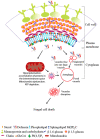
Model of the proposed mechanisms of action of Ppdef1. Ppdef1 binds to the fungal cell wall and is endocytosed from the plasma membrane. It penetrates the cytoplasm and targets mitochondria, causing the hyperpolarisation of the mitochondrial membrane potential and ROS, induces programmed cell death, induces vacuolar fusion and causes fungal cell death. This model is an addition to the previously described mechanisms of action of defensins [8].

Similar articles

Cited by

References

    1. Marmiroli N., Maestri E. Plant peptides in defense and signaling. Peptides. 2014;56:30–44. doi: 10.1016/j.peptides.2014.03.013. - DOI - PubMed
    1. De Beer A., Vivier M.A. Four plant defensins from an indigenous South African Brassicaceae species display divergent activities against two test pathogens despite high sequence similarity in the encoding genes. BMC Res. Notes. 2011;4:459. doi: 10.1186/1756-0500-4-459. - DOI - PMC - PubMed
    1. de Oliveira Carvalho A., Moreira Gomes V. Plant defensins and defensin-like peptides-biological activities and biotechnological applications. Curr. Pharm. Des. 2011;17:4270–4293. doi: 10.2174/138161211798999447. - DOI - PubMed
    1. Lay F., Anderson M. Defensins-components of the innate immune system in plants. Curr. Protein Pept. Sci. 2005;6:85–101. doi: 10.2174/1389203053027575. - DOI - PubMed
    1. García-Olmedo F., Molina A., Alamillo J.M., Rodríguez-Palenzuéla P. Plant defense peptides. Pept. Sci. 1998;47:479–491. doi: 10.1002/(SICI)1097-0282(1998)47:6<479::AID-BIP6>3.0.CO;2-K. - DOI - PubMed

LinkOut - more resources