. 2024 Jan 16;10(1):72.
doi: 10.3390/jof10010072.

Molecular Signaling and Metabolic Responses during the Interaction between Human Keratinocytes (HaCaT) and the Dermatophyte Trichophyton rubrum

Affiliations

Molecular Signaling and Metabolic Responses during the Interaction between Human Keratinocytes (HaCaT) and the Dermatophyte Trichophyton rubrum

Monise Fazolin Petrucelli et al. J Fungi (Basel). .

Abstract

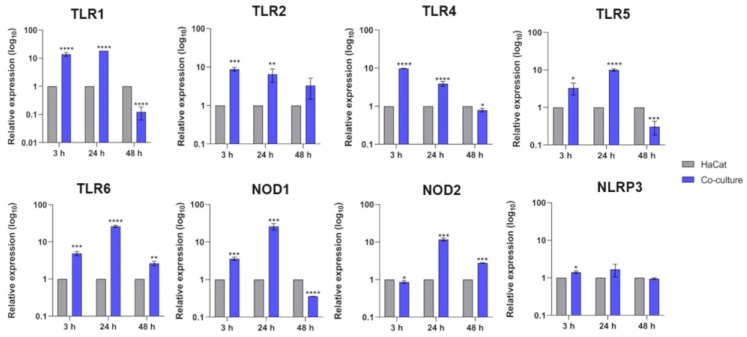
Trichophyton rubrum is the leading causative agent of dermatophytosis worldwide. Keratinocytes are the first line of defense that drives an immune response against fungal invasion. Host-specific pattern recognition receptors (PRRs) recognize pathogen-associated molecular patterns (PAMPs) to trigger immunological pathways. Fungal cell wall components are the primary sources of fungal PAMPs, and some pathogens increase cell wall rearrangement to evade the immune system. Glycolysis and enhanced lactate levels are critical for improving host immune responses to fungal infections. Using reverse transcription-quantitative polymerase chain reaction (RT-qPCR), we evaluated the transcriptional responses of human genes involved in fungal recognition and glycolytic metabolism and fungal cell-wall-related genes in a co-culture model of human keratinocytes with T. rubrum. We observed the upregulation of several Toll-like receptors (TLRs), NOD-like receptors (NLRs), and glycolytic genes. Complementarily, we measured intra- and extracellular glucose levels and the increase in lactate production in the co-culture supernatant. We noted a distinct transcriptional regulation pattern of fungal cell-wall-related genes from fungal growth on keratin as the primary carbon source compared to co-culture with human keratinocytes. Our results showed new insights into the transcriptional adaptation of keratinocytes, particularly in regulating genes involved in sensing and metabolic processes, during the interaction with T. rubrum.

Keywords: PRRs; Trichophyton rubrum; cell wall; glucose; immune response; keratinocytes; lactate; pathogen–host interaction.


Conflict of interest statement

The authors declare that the research was conducted in the absence of any commercial or financial relationships that could be construed as potential conflicts of interest.

Figures

Figure 1
Figure 1
Relative expression analyses of human keratinocytes’ (HaCaT) PRRs (TLRs and NLRs) during co-culture with T. rubrum. Statistical significance was determined using an unpaired Student’s t-test with Holm–Sidak correction for multiple testing considering * p < 0.05, ** p < 0.01, *** p < 0.001, and **** p < 0.0001.
Figure 2
Figure 2
Relative expression analyses of human genes involved in nutrient signaling and glycolytic metabolism during co-culture with T. rubrum. Statistical significance was determined using an unpaired Student’s t-test with Holm–Sidak correction for multiple testing considering * p < 0.05, ** p < 0.01, *** p < 0.001, and **** p < 0.0001.
Figure 3
Figure 3
Intracellular (A) and extracellular (B) glucose content comparison between HaCaT keratinocytes (control) and HaCaT keratinocytes co-cultured with T. rubrum. Statistical significance was determined using an unpaired Student’s t-test with Holm–Sidak correction for multiple testing considering * p < 0.05 and *** p < 0.001. N/D means the absence of significant glucose levels detected in the control and co-culture conditions.
Figure 4
Figure 4
Extracellular lactate content in HaCaT keratinocytes (control) and HaCaT keratinocytes co-cultured with T. rubrum. Statistical significance was determined using an unpaired Student’s t-test with Holm–Sidak correction for multiple testing considering ** p < 0.01 and *** p < 0.001.
Figure 5
Figure 5
Intracellular (A) and extracellular (B) LDH activity measurements in HaCaT keratinocytes (control) and HaCaT keratinocytes co-cultured with T. rubrum. Statistical significance was determined using an unpaired Student’s t-test with Holm–Sidak correction for multiple testing considering ** p < 0.01 and *** p < 0.001. N/D means the absence of significant LDH activity detected in the control and co-culture conditions.
Figure 6
Figure 6
Relative expression analysis of fungal cell wall genes during co-culture with keratinocytes. Expression levels for each condition are compared to the control (T. rubrum grown in RPMI-1640 medium without HaCaT cells). Statistical significance was determined using an unpaired Student’s t-test with Holm–Sidak correction for multiple testing considering * p < 0.05, ** p < 0.01, *** p < 0.001, and **** p < 0.0001.
Figure 7
Figure 7
Relative expression analysis of fungal cell-wall-related genes during switching from glucose to keratin. Expression levels for each condition are compared to the control (T. rubrum grown in glucose medium). Statistical significance was determined using an unpaired Student’s t-test with Holm–Sidak correction for multiple testing considering *** p < 0.001 and **** p < 0.0001.
Figure 8
Figure 8
Proposed model of early host–pathogen interaction focusing on the signaling of human keratinocytes co-cultured with T. rubrum. Upon fungal recognition by keratinocytes, immunological cascades are triggered to prevent fungal colonization. TLRs mediate the recognition of fungal PAMPs and activate transcription responses enacted by AP-1 and NF-κB. At the same time, the PI3K/Akt/mTOR pathway controls glucose metabolism, which also results in NF-κB recruitment. In addition, HIF-1α plays a role in pyruvate dehydrogenase (PHDA), lactate dehydrogenase (LDHA), and glucose transporter (glut) expressions. The Warburg effect might also result from glucose metabolism after fungal interaction with HaCaT cells, through the monocarboxylate transporter MCL in the keratinocyte membrane. Fungus contacting HaCaT cells triggers the expression of enzymes involved in the biosynthesis of cell wall components. We have created this figure with the help of BioRender.com.

References

    1. Petrucelli M.F., de Abreu M.H., Cantelli B.A.M., Segura G.G., Nishimura F.G., Bitencourt T.A., Marins M., Fachin A.L. Epidemiology and diagnostic perspectives of dermatophytoses. J. Fungi. 2020;6:310. doi: 10.3390/jof6040310. - DOI - PMC - PubMed
    1. Martinez-Rossi N.M., Peres N.T.A., Bitencourt T.A., Martins M.P., Rossi A. State-of-the-Art dermatophyte infections: Epidemiology aspects, pathophysiology, and resistance mechanisms. J. Fungi. 2021;7:629. doi: 10.3390/jof7080629. - DOI - PMC - PubMed
    1. Ciesielska A., Kawa A., Kanarek K., Soboń A., Szewczyk R. Metabolomic analysis of Trichophyton rubrum and Microsporum canis during keratin degradation. Sci. Rep. 2021;11:3959. doi: 10.1038/s41598-021-83632-z. - DOI - PMC - PubMed
    1. Cruz A.H.S., Santos R.S., Martins M.P., Peres N.T.A., Trevisan G.L., Mendes N.S., Martinez-Rossi N.M., Rossi A. Relevance of nutrient-sensing in the pathogenesis of Trichophyton rubrum and Trichophyton interdigitale. Front. Fungal Biol. 2022;3:858968. doi: 10.3389/ffunb.2022.858968. - DOI - PMC - PubMed
    1. Martins-Santana L., Petrucelli M.F., Sanches P.R., Martinez-Rossi N.M., Rossi A. Peptidase regulation in Trichophyton rubrum is mediated by the synergism between alternative splicing and StuA-dependent transcriptional mechanisms. Front. Microbiol. 2022;13:2251. doi: 10.3389/fmicb.2022.930398. - DOI - PMC - PubMed

LinkOut - more resources