

The Microscopic Morphology of Mouthparts and Their Sensilla in the Mycophagous Ladybeetle Illeis chinensis (Coleoptera: Coccinellidae)
- PMID: 38249052
- PMCID: PMC10816638
- DOI: 10.3390/insects15010046
The Microscopic Morphology of Mouthparts and Their Sensilla in the Mycophagous Ladybeetle Illeis chinensis (Coleoptera: Coccinellidae)
Abstract
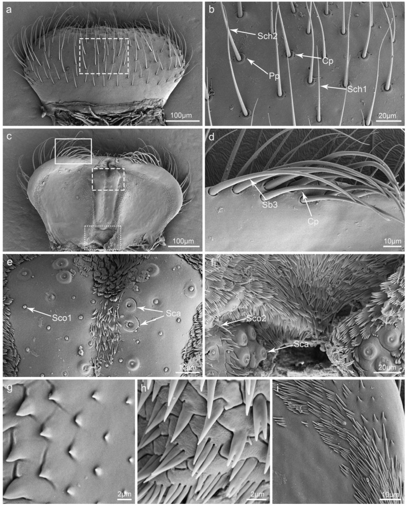
The morphological diversity of insect mouthparts is closely related to changes in food sources and diets. Research into the structures of insect mouthparts may help to establish a fundamental basis for a better understanding of insect feeding mechanisms. In this study, we examined the fine morphology of the mouthparts of Illeis chinensis using scanning electron microscopy. We paid particular attention to the types, quantities, and distribution of sensilla on the mouthparts. Our results showed that the basic components of the mouthparts of I. chinensis are the same as those in other lady beetles, i.e., the labrum, mandible, maxillae, labium, and hypopharynx. We also found structural specialization indicating adaptation to fungal feeding. On the mouthparts, there are eight kinds of sensilla and two kinds of glandular structures, including sensilla chaetica, sensilla basiconica, sensilla styloconica, sensilla coeloconica, sensilla campaniformia, sensilla placodea, sensilla digitiformia, Böhm bristles, perforated plates, and cuticular pores. This is the first time that sensilla digitiformia has been reported in ladybirds. Finally, variations in mouthparts among ladybirds with differing diets, as well as the putative functions of each of the mouthparts and sensilla, were discussed. This research can provide a reference for understanding the functions of the mouthparts in ladybird feeding behavior and thereby contribute to the development of precise insect behavior regulation and management strategies.
Keywords: Illeis chinensis; fine morphology; mouthparts; scanning electron microscopy; sensilla.
Conflict of interest statement
The authors declare no conflicts of interest.
Figures


Similar articles
-
Functional morphology of the mouthparts of lady beetle Coccinella transversoguttata (Coccinellidae, Coleoptera), with reference to their feeding mechanism.J Morphol. 2019 May;280(5):701-711. doi: 10.1002/jmor.20976. Epub 2019 Mar 22. J Morphol. 2019. PMID: 30901106
-
A SEM study of the antenna and mouthparts of Omosita colon (Linnaeus) (Coleoptera: Nitidulidae).Microsc Res Tech. 2016 Dec;79(12):1152-1164. doi: 10.1002/jemt.22770. Epub 2016 Sep 15. Microsc Res Tech. 2016. PMID: 27629391
-
Morphology of the Mouthparts of Ladybeetle Vibidia duodecimguttata (Coleoptera: Coccinellidae), with Emphasis on Their Sensilla.Insects. 2024 Oct 31;15(11):854. doi: 10.3390/insects15110854. Insects. 2024. PMID: 39590452 Free PMC article.
-
Morphology of Mouthparts and Distribution of Sensilla in Immature Stages and Adults of Parthenium Beetles.Microsc Microanal. 2025 May 9;31(3):ozaf035. doi: 10.1093/mam/ozaf035. Microsc Microanal. 2025. PMID: 40358597
-
The Right Tool for the Job: A Review of Insect Mouthparts as a Tool Kit for Biomimetic Studies.Biomimetics (Basel). 2025 Mar 24;10(4):196. doi: 10.3390/biomimetics10040196. Biomimetics (Basel). 2025. PMID: 40277595 Free PMC article. Review.
References
-
- Betz O., Thayer M.K., Newton A.F. Comparative morphology and evolutionary pathways of the mouthparts in spore-feeding Staphylinoidea (Coleoptera) Acta Zool. 2003;84:179–238. doi: 10.1046/j.1463-6395.2003.00147.x. - DOI
-
- Samways M.J., Osborn R., Saunders T.S. Mandibles form relative to the main food type in ladybirds (Coleoptera: Coccinellidae) Biocontrol Sci. Technol. 1997;7:275–286. doi: 10.1080/09583159730974. - DOI
Grants and funding
- 32160120, 32270468/National Natural Science Foundation of China
- 2022YFC2601200/National Key R&D Program of China
- NABRI202203/the Project of the Northeast Asia Biodiversity Research Center
- 21JR7RA848/the Youth Natural Science Foundation of Gansu Province
- Gaufx‑03Y05/the Funds for Fuxi Young Scientific Talents of Gansu Agricultural University
LinkOut - more resources
Full Text Sources
