Review
. 2023 Dec 21;15(12):e50899.
doi: 10.7759/cureus.50899. eCollection 2023 Dec.

Can Echocardiography Aid in Diagnosing Vascular Rings?

Affiliations
Review

Can Echocardiography Aid in Diagnosing Vascular Rings?

Yasser A Bhat et al. Cureus. .

Abstract

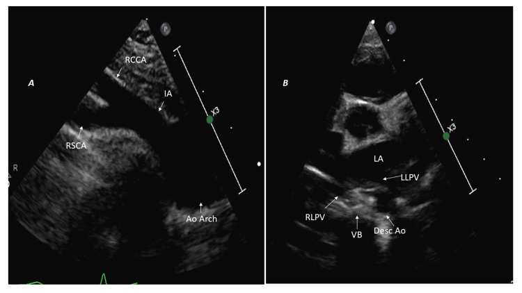
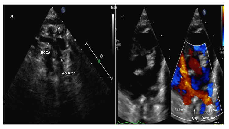
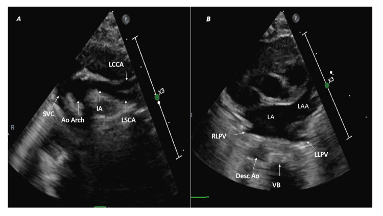
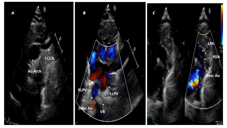
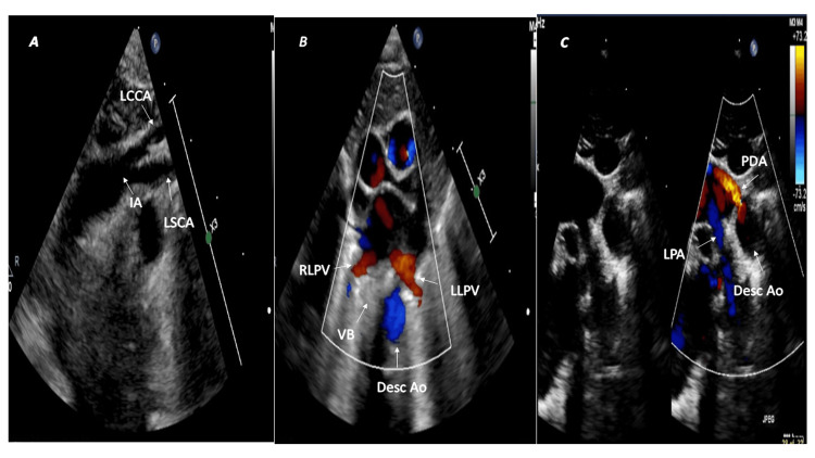
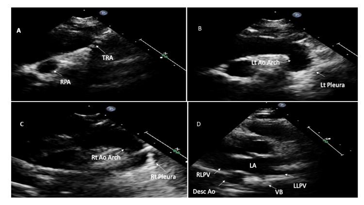
Even though cardiac computed tomography and magnetic resonance imaging are the gold standard for evaluating the aortic arch in the context of vascular rings in children, echocardiography is usually the first-line modality. The echocardiographic evaluation of the aortic arch in the context of vascular rings in children has received little attention. This article details the step-by-step echocardiographic assessment of the aortic arch in vascular ring patients.

Keywords: arch anomalies; circumflex arch; double aortic arch; echocardiography; vascular ring.


Conflict of interest statement

The authors unequivocally state that all figures, comprising of pictures and videos, have been collected exclusively from their own patients.

Figures

Figure 1
Figure 1. Left aortic arch with normal branching pattern and left-sided descending aorta
(A) Suprasternal short-axis view: first branch (IA) courses toward the right and bifurcates. (B) High parasternal short-axis image: left-sided descending aorta (Desc Ao) located posterior to left lower pulmonary vein (LLPV) on the left side of the vertebral body (VB). Ao Arch: aortic arch; IA: innominate artery; RSCA: right subclavian artery; RCCA: right common carotid artery; RLPV: right lower pulmonary vein; LA: left atrium.
Figure 2
Figure 2. Left aortic arch with aberrant right subclavian artery
(A) Suprasternal short-axis view shows the first branch (RCCA) courses toward the right but does not bifurcate. (B) High parasternal short-axis color comparing image: left-sided descending aorta (Desc Ao) located posterior to left lower pulmonary vein (LLPV) on the left side of the vertebral body (VB). Ao Arch: aortic arch; IV: innominate vein; RCCA: right common carotid artery; RLPV: right lower pulmonary vein.
Figure 3
Figure 3. Right aortic arch with mirror branching pattern and right-sided descending aorta
(A) Suprasternal short-axis view: first branch (IA) courses toward the left and bifurcates. (B) High parasternal short-axis image: right-sided descending aorta (Desc Ao) located posterior to right lower pulmonary vein (RLPV) on the right side of the vertebral body (VB). Ao Arch: aortic arch; IA: innominate artery; LSCA: left subclavian artery; LCCA: left common carotid artery; LLPV: left lower pulmonary vein; LA: left atrium; LAA: left atrial appendage.
Figure 4
Figure 4. Right aortic arch with aberrant left subclavian artery and left-sided patent ductus arteriosus
(A) The first branch (LCCA) courses leftwards but does not bifurcate. (B) The right descending aorta (Desc Ao) is posterior to the right lower pulmonary vein (RLPV) on the right side of the vertebral body (VB). (C) A tiny left-sided patent ductus arteriosus (PDA) completes the vascular ring. Ao Arch: aortic arch; LCCA: left common carotid artery; LLPV: left lower pulmonary vein; LPA: left pulmonary artery.
Figure 5
Figure 5. Right circumflex aorta and left patent ductus arteriosus
(A) Right aortic arch with mirror image branching pattern. The left innominate artery (IA) courses leftwards and bifurcates into the left subclavian artery (LSCA) and left common carotid artery (LCCA). (B) The left descending thoracic aorta (Desc Ao) lies posterior to the left lower pulmonary vein (LLPV) on the left side of the vertebral body (VB). (C) The left patent ductus arteriosus (PDA) completes the vascular ring. RLPV: right lower pulmonary vein; LPA: left pulmonary artery.
Figure 6
Figure 6. Sequential two-dimensional echocardiographic images of double aortic arch
(A) Suprasternal long axis sweep with the probe position at 12 o'clock shows tracheal cartilages (TRA). (B) Tilting the probe leftwards shows the left aortic arch (Lt Ao Arch) and left pleura (Lt pleura). (C) Tilting the probe rightwards from 12 o'clock shows the right aortic arch (Rt Ao Arch) and right pleura (Rt pleura). (D) Right descending thoracic aorta (Desc Ao) in this patient with double aortic arch. RPA: right pulmonary artery; VB: vertebral body; RLPV: right lower pulmonary vein; LLPV: left lower pulmonary vein; RPA: right pulmonary artery; LA: left atrium.

References

    1. Is there a role for echocardiography in vascular ring diagnosis? Snarr BS, Dyer A, Thankavel PP. J Am Soc Echocardiogr. 2018;31:965–966. - PubMed
    1. Morphology of vascular ring arch anomalies influences prognosis and management. Ctori E, Crucean A, Pinkey B, et al. Arch Dis Child. 2020;106:477–483. - PubMed
    1. Congenital variants and anomalies of the aortic arch. Hanneman K, Newman B, Chan F. Radiographics. 2017;37:32–51. - PubMed
    1. Echocardiography in the preoperative evaluation of vascular rings. Lillehei CW, Colan S. J Pediatr Surg. 1992;27:1118–1120. - PubMed
    1. Aortic uncrossing and tracheobronchopexy corrects tracheal compression and tracheobronchomalacia associated with circumflex aortic arch. Kamran A, Friedman KG, Jennings RW, Baird CW. J Thorac Cardiovasc Surg. 2020;160:796–804. - PubMed

LinkOut - more resources