

How Kirschner Wires Crossing Each Other at the Fracture Site Affect Radiological and Clinical Results in Children With Gartland Type 3 Supracondylar Humerus Fractures?
- PMID: 38249197
- PMCID: PMC10799975
- DOI: 10.7759/cureus.50919
How Kirschner Wires Crossing Each Other at the Fracture Site Affect Radiological and Clinical Results in Children With Gartland Type 3 Supracondylar Humerus Fractures?
Abstract
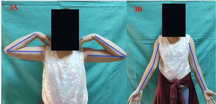
Background In this study, we compared two groups of children with Gartland Type 3 supracondylar humerus fractures with respect to the crossing point of Kirschner wires (K-wires) in terms of radiological and clinical results after closed reduction and fixation with the crossed-pin technique. We hypothesized that even if medial and lateral pins cross each other at the fracture line, satisfactory radiological and clinical results would be achieved with the crossed-pin technique. Methodology A total of 59 patients with Gartland extension Type 3 supracondylar humerus fractures who underwent closed reduction and percutaneous crossed-pin fixation were included in the study. K-wires were crossing each other proximal to the fracture site in the proximal crossing group and at the fracture level in the fracture site crossing group. Loss of reduction, Baumann angle, shaft condyle angle, range of motion, and carrying angle were compared between the two groups. Results There were 43 males and 16 females in this study, with a mean age of 5.3±2.4 years. The average follow-up duration was 21.9 ± 5.2 weeks. In terms of loss of reduction in the coronal and sagittal planes, there was no statistical difference between the two groups. When the Baumann angle and shaft condylar angle of both groups were analyzed, no statistically significant differences were found at both early postoperative examination and final follow-up. Conclusions Although the crossing point of K-wires has been shown to be an important factor in the protection of reduction in biomechanical studies, it was not a significant factor for loss of reduction in this study. Factors except for the crossing point of K-wires may play a more important role in the outcomes of crossed-pin fixation.
Keywords: closed reduction; crossed-pin fixation; fracture line; gartland type 3 fractures; supracondylar humerus fracture.
Copyright © 2023, Kilic et al.
Conflict of interest statement
The authors have declared that no competing interests exist.
Figures


Similar articles
-
[Short-term effectiveness of transverse antecubital incision for failed closed reduction of Gartland type Ⅲ supracondylar humerus fractures in children].Zhongguo Xiu Fu Chong Jian Wai Ke Za Zhi. 2023 May 15;37(5):566-571. doi: 10.7507/1002-1892.202211033. Zhongguo Xiu Fu Chong Jian Wai Ke Za Zhi. 2023. PMID: 37190833 Free PMC article. Chinese.
-
Operative treatment of supracondylar fractures of the humerus in children. The consequences of pin placement.J Bone Joint Surg Am. 2001 May;83(5):735-40. J Bone Joint Surg Am. 2001. PMID: 11379744
-
The posterior intrafocal pin improves sagittal alignment in Gartland type III paediatric supracondylar humeral fractures.Injury. 2016 Apr;47(4):842-7. doi: 10.1016/j.injury.2015.12.031. Epub 2015 Dec 31. Injury. 2016. PMID: 26777466
-
Biomechanical analysis of supracondylar humerus fracture pinning for fractures with coronal lateral obliquity.J Pediatr Orthop. 2012 Mar;32(2):196-200. doi: 10.1097/BPO.0b013e318242a99a. J Pediatr Orthop. 2012. PMID: 22327455
-
Low incidence of ulnar nerve injury with crossed pin placement for pediatric supracondylar humerus fractures using a mini-open technique.J Orthop Trauma. 2005 Mar;19(3):158-63. doi: 10.1097/00005131-200503000-00002. J Orthop Trauma. 2005. PMID: 15758668 Review.
Cited by
-
Objective Fluoroscopic Image-Based Assessment of Intraoperative Wire Navigation Skill Agrees with Subjective Expert Opinion.Iowa Orthop J. 2025;45(1):49-59. Iowa Orthop J. 2025. PMID: 40606727 Free PMC article.
References
-
- Management of supracondylar humerus fractures in children: current concepts. Abzug JM, Herman MJ. J Am Acad Orthop Surg. 2012;20:69–77. - PubMed
-
- Supracondylar fractures of the humerus in children. Otsuka NY, Kasser JR. J Am Acad Orthop Surg. 1997;5:19–26. - PubMed
-
- A 10-year study of the changes in the pattern and treatment of 6,493 fractures. Cheng JC, Ng BK, Ying SY, Lam PK. https://journals.lww.com/pedorthopaedics/abstract/1999/05000/a_10_year_s.... J Pediatr Orthop. 1999;19:344–350. - PubMed
-
- The treatment of pediatric supracondylar humerus fractures. Howard A, Mulpuri K, Abel MF, et al. J Am Acad Orthop Surg. 2012;20:320–327. - PubMed
-
- The displaced supracondylar humerus fracture: indications for surgery and surgical options: a 2014 update. Ladenhauf HN, Schaffert M, Bauer J. Curr Opin Pediatr. 2014;26:64–69. - PubMed
LinkOut - more resources
Full Text Sources