

The function of previously unappreciated exerkines secreted by muscle in regulation of neurodegenerative diseases
- PMID: 38249295
- PMCID: PMC10796786
- DOI: 10.3389/fnmol.2023.1305208
The function of previously unappreciated exerkines secreted by muscle in regulation of neurodegenerative diseases
Abstract
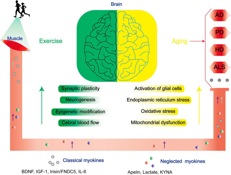
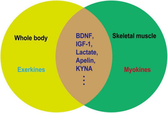
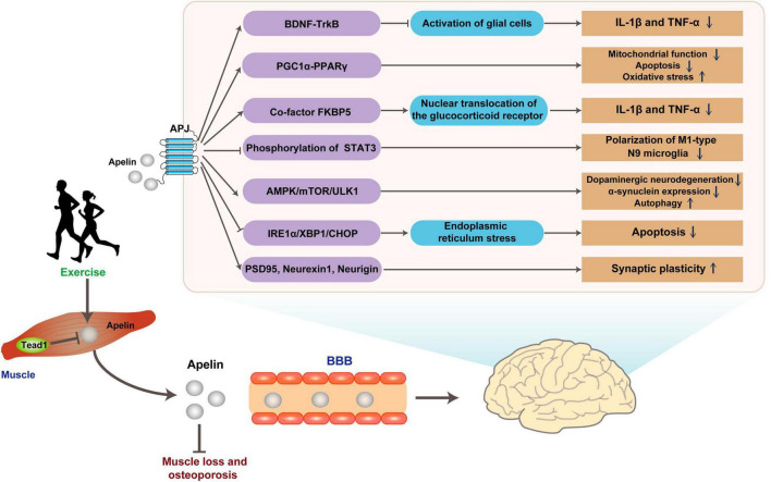
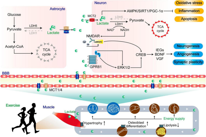
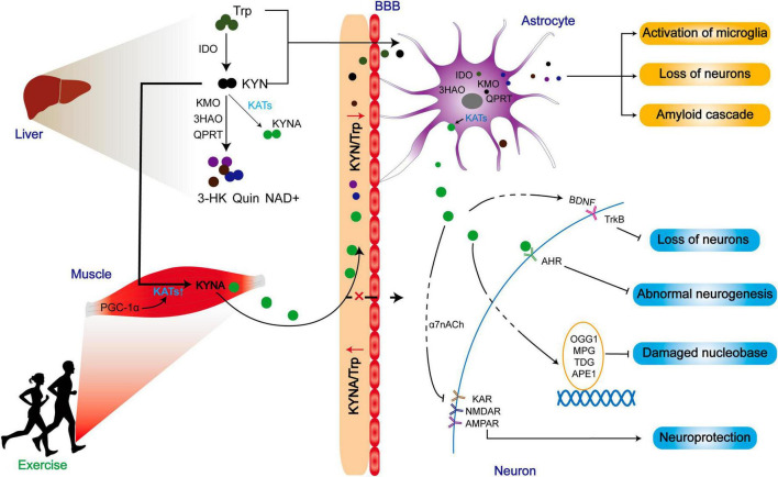
The initiation and progression of neurodegenerative diseases (NDs), distinguished by compromised nervous system integrity, profoundly disrupt the quality of life of patients, concurrently exerting a considerable strain on both the economy and the social healthcare infrastructure. Exercise has demonstrated its potential as both an effective preventive intervention and a rehabilitation approach among the emerging therapeutics targeting NDs. As the largest secretory organ, skeletal muscle possesses the capacity to secrete myokines, and these myokines can partially improve the prognosis of NDs by mediating the muscle-brain axis. Besides the well-studied exerkines, which are secreted by skeletal muscle during exercise that pivotally exert their beneficial function, the physiological function of novel exerkines, e.g., apelin, kynurenic acid (KYNA), and lactate have been underappreciated previously. Herein, this review discusses the roles of these novel exerkines and their mechanisms in regulating the progression and improvement of NDs, especially the significance of their functions in improving NDs' prognoses through exercise. Furthermore, several myokines with potential implications in ameliorating ND progression are proposed as the future direction for investigation. Elucidation of the function of exerkines secreted by skeletal muscle in the regulation of NDs advances the understanding of its pathogenesis and facilitates the development of therapeutics that intervene in these processes to cure NDs.
Keywords: KYNA; apelin; exercise; lactate; myokine; neurodegenerative disease.
Copyright © 2024 Bian, Wang, Wang and Lou.
Conflict of interest statement
The authors declare that the research was conducted in the absence of any commercial or financial relationships that could be construed as a potential conflict of interest.
Figures
Similar articles
-
Physical Exercise-Induced Myokines in Neurodegenerative Diseases.Int J Mol Sci. 2021 May 28;22(11):5795. doi: 10.3390/ijms22115795. Int J Mol Sci. 2021. PMID: 34071457 Free PMC article. Review.
-
Exercise-Induced Myokines With Therapeutic Potential for Muscle Wasting.Front Physiol. 2019 Mar 29;10:287. doi: 10.3389/fphys.2019.00287. eCollection 2019. Front Physiol. 2019. PMID: 30984014 Free PMC article.
-
Exerkines and osteoarthritis.Front Physiol. 2023 Dec 1;14:1302769. doi: 10.3389/fphys.2023.1302769. eCollection 2023. Front Physiol. 2023. PMID: 38107476 Free PMC article. Review.
-
The role of exerkines on brain mitochondria: a mini-review.J Appl Physiol (1985). 2023 Jan 1;134(1):28-35. doi: 10.1152/japplphysiol.00565.2022. Epub 2022 Nov 23. J Appl Physiol (1985). 2023. PMID: 36417200 Free PMC article. Review.
-
Forgot to Exercise? Exercise Derived Circulating Myokines in Alzheimer's Disease: A Perspective.Front Neurol. 2021 Jun 30;12:649452. doi: 10.3389/fneur.2021.649452. eCollection 2021. Front Neurol. 2021. PMID: 34276532 Free PMC article. Review.
Cited by
-
Protective role of coenzyme Q10 against trihexyphenidyl-induced pulmonary toxicity in Wistar rats.BMC Pharmacol Toxicol. 2025 May 31;26(1):114. doi: 10.1186/s40360-025-00955-7. BMC Pharmacol Toxicol. 2025. PMID: 40450364 Free PMC article.
-
Oxidative Stress in Neurodegenerative Disorders: A Key Driver in Impairing Skeletal Muscle Health.Int J Mol Sci. 2025 Jun 16;26(12):5782. doi: 10.3390/ijms26125782. Int J Mol Sci. 2025. PMID: 40565245 Free PMC article. Review.
-
'Exerkines': A Comprehensive Term for the Factors Produced in Response to Exercise.Biomedicines. 2024 Sep 1;12(9):1975. doi: 10.3390/biomedicines12091975. Biomedicines. 2024. PMID: 39335489 Free PMC article. Review.
-
From Brain to Muscle: The Role of Muscle Tissue in Neurodegenerative Disorders.Biology (Basel). 2024 Sep 12;13(9):719. doi: 10.3390/biology13090719. Biology (Basel). 2024. PMID: 39336146 Free PMC article. Review.
-
The Biology and Biochemistry of Kynurenic Acid, a Potential Nutraceutical with Multiple Biological Effects.Int J Mol Sci. 2024 Aug 21;25(16):9082. doi: 10.3390/ijms25169082. Int J Mol Sci. 2024. PMID: 39201768 Free PMC article. Review.
References
-
- Akyüz M., Doğru Y., Nalcakan G. R., Ulman C., Taş M., Varol R. (2021). The effects of various strength training intensities on blood cardiovascular risk markers in healthy men. Turk. J. Biochem. 46 693–701.
Publication types
LinkOut - more resources
Full Text Sources
Research Materials