Review
. 2023 Dec 30:24:100939.
doi: 10.1016/j.mtbio.2023.100939. eCollection 2024 Feb.

Enhancing the mechanical strength of 3D printed GelMA for soft tissue engineering applications

Affiliations
Review

Enhancing the mechanical strength of 3D printed GelMA for soft tissue engineering applications

Ao Guo et al. Mater Today Bio. .

Abstract

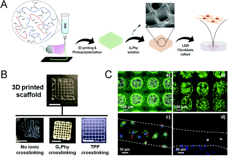
Gelatin methacrylate (GelMA) hydrogels have gained significant traction in diverse tissue engineering applications through the utilization of 3D printing technology. As an artificial hydrogel possessing remarkable processability, GelMA has emerged as a pioneering material in the advancement of tissue engineering due to its exceptional biocompatibility and degradability. The integration of 3D printing technology facilitates the precise arrangement of cells and hydrogel materials, thereby enabling the creation of in vitro models that simulate artificial tissues suitable for transplantation. Consequently, the potential applications of GelMA in tissue engineering are further expanded. In tissue engineering applications, the mechanical properties of GelMA are often modified to overcome the hydrogel material's inherent mechanical strength limitations. This review provides a comprehensive overview of recent advancements in enhancing the mechanical properties of GelMA at the monomer, micron, and nano scales. Additionally, the diverse applications of GelMA in soft tissue engineering via 3D printing are emphasized. Furthermore, the potential opportunities and obstacles that GelMA may encounter in the field of tissue engineering are discussed. It is our contention that through ongoing technological progress, GelMA hydrogels with enhanced mechanical strength can be successfully fabricated, leading to the production of superior biological scaffolds with increased efficacy for tissue engineering purposes.


Conflict of interest statement

The authors declare that they have no known competing financial interests or personal relationships that could have appeared to influence the work reported in this paper.

Figures

Image 1
Graphical abstract
Fig. 1
Fig. 1
A) Schematic representation of the formation of GelMA from gelatin by substitution reaction. B) Modification Methods and Tissue Engineering Applications of GelMA Hydrogel. C) Schematic diagram of the formation of IPNs.
Fig. 2
Fig. 2
Reinforcement of hydrogel scaffolds by a network of IPNs. A) The SEM images of the porosity of hydrogel scaffolds with different HA concentration ratios. B) False-colour SEM image of adherent cells on the surface of the scaffold after seeding C28 cells onto the surface of the sample and incubating the scaffold for 24 h after fixation. Reproduced with permission [79]. Copyright 2021, Elsevier. C) Illustration of preparing alginate/alginate sulfateGelMA IPN bioink. D) Schematic of crosslinking processes. The constructs were either cultured in vitro for 6 weeks or subcutaneously implanted for 4 weeks. E) is the hydrogel pattern printed by different bio-inks. F) shows the representative images of live/dead staining of MSCs within constructs at various time points (day 1, 21, and 42) throughout the culture duration. G) Cell survival to 3D printed constructs. Reproduced with permission [80]. Copyright 2021, Elsevier.
Fig. 3
Fig. 3
A) Reinforcement of GelMA/chitosan 3D polymer scaffolds with ionic crosslinking agent (G1Phy). B) Visual examination and structural integrity changes that a photochemically 3D printed scaffold (four layers) immediately experienced after immersion in distilled water containing or not the G1Phy or TPP. Scale bar is 0.5 cm. C) Live/dead assay on G1Phy (a) and TPP (b) scaffolds after 24 h of incubation. Pictures were taken of scaffold strands; confocal immunostaining assay performed on G1Phy (c) and TPP (d) scaffolds after 24 h of incubation. Reproduced with permission [90]. Copyright 2020, Royal society of chemistry.
Fig. 4
Fig. 4
A) Scanning electron microscopic images of lyophilized GelMA, GelMA–dhECM, GelMA–MeHA and GelMA–MeHA–dhECM hydrogels with mTGase treatment. B) Brightfield images of 3D printed 2 layered-grid constructs using these four hydrogels. C) Cell viability analysis of the printed cell-laden hydrogels prepared using these four hydrogels. Reproduced with permission [86]. Copyright 2021, Multidisciplinary Digital Publishing Institute. D) Viability staining of 3D bioprinted GelMA:HAMA (1:1, 2:1 and 3:1) constructs over days 0, 1 and 7. E) Day 0 storage moduli of DoE generated GelMA:HAMA mixtures. (i): 3D surface plot of storage moduli vs crosslinking and material mixtures. (ii): Corresponding normal probability plot of the residuals. Reproduced with permission [88]. Copyright 2022, Elsevier.
Fig. 5
Fig. 5
Interpenetrating networks (IPNs) of calcium alginate-GelMA fortified by (BNC). A) The schematic illustration of 3D-bioprinting bioink production. B) (i): Printing model of the 2.5 % GelMA group, (ii): Printing model of the 5 % GelMA group, (iii): Printing model of the 10 % GelMA group, (iv): Printing model of the 5 % GelMA +0.3 % BNC group (scale bar = 1 mm). C) (i): Electrical conductivity, (ii): Compressive modulus. D) Cytoskeletal staining of RSC96 cytoskeleton on different hydrogel scaffolds on days 4 and 7 (White line in Day 7 shows the outline of the scaffold, and the arrow marks the direction of the long axis of the scaffold). E) Live/dead cell staining: The viability of RSC96 cells in the 3D-bioprinted scaffold within 7 days of culture. F) In vivo experiment: HE and S100β immunohistochemical staining of paraffin embedded sections at 2 and 4 weeks (Scale bar 50 μm). Reproduced with permission [82]. Copyright 2021, Elsevier.
Fig. 6
Fig. 6
A) The schematic illustration of cell-laden GelMA-alginate core-shell microcapsules fabricated using coaxial electrostatic microdroplet system and their application in endodontic regeneration. B) Highermagnification optical images of the core-shell microcapsules and the GelMA microgels and the corresponding SEM images after lyophilization. The scale bars in i, ii, iv and v are 200 μm. The scale bars in iii, vi are 50 μm. C) H&E and immunohistochemical stained images of paraffin sections from each group under light microscope. (i): Acellular GelMA microgels group (group G), (ii): hDPSCs-laden GelMA microgels group (group DP), (iii): GelMA microgels laden with 3:1 hDPSCs: HUVECs group (group 3:1), and (iv): GelMA microgels laden with 1:1 hDPSCs: HUVECs group (group 1:1). Scale bars in the first, second, third and fourth columns were 10 00, 10 0, 50, and 20 μm, respctively. Reproduced with permission [193]. Copyright 2022, Elsevier.
Fig. 7
Fig. 7
A) Scheme representation of the synthesize of Gel-NH2 and Gel-SH. B) 1H NMR spectra of gelatin, Gel-NH2, Gel-SH (one-step) and Gel-SH (two-steps) confirming the chemical grafting of thiol on the gelatin structure. C) FTIR spectra of gelatin, Gel-NH2 and Gel-SH confirming the thiolation process. D) The photographic images of gelatin based hydrogel, i before and ii after crosslinking with visible light. E) Compressive stress-strain curves. F) Compressive strength, G) elastic modulus, H) tensile strength, I) toughness and J) elongation changes as a function of PD-LAP, with the data shown as means ± SD (n = 3) (*: P < 0.05). Reproduced with permission [194]. Copyright 2020, Elsevier.

Cited by

References

    1. Lei L., Bai Y., Qin X., Liu J., Huang W., Lv Q. Current Understanding of hydrogel for drug release and tissue engineering. Gels. 2022;8:301. doi: 10.3390/gels8050301. - DOI - PMC - PubMed
    1. Zhong H., Huang J., Wu J., Du J. Electrospinning nanofibers to 1D, 2D, and 3D scaffolds and their biomedical applications. Nano Res. 2022;15:787–804. doi: 10.1007/s12274-021-3593-7. - DOI
    1. Levato R., Jungst T., Scheuring R.G., Blunk T., Groll J., Malda J. From shape to function: the next step in bioprinting. Adv. Mater. 2020;32 doi: 10.1002/adma.201906423. - DOI - PMC - PubMed
    1. Xiao S., Zhao T., Wang J., Wang C., Du J., Ying L., Lin J., Zhang C., Hu W., Wang L., Xu K. Gelatin methacrylate (GelMA)-Based hydrogels for cell transplantation: an effective strategy for tissue engineering. Stem Cell Rev and Rep. 2019;15:664–679. doi: 10.1007/s12015-019-09893-4. - DOI - PubMed
    1. Morgan F.L.C., Moroni L., Baker M.B. Dynamic bioinks to advance bioprinting. Adv. Healthcare Mater. 2020;9 doi: 10.1002/adhm.201901798. - DOI - PubMed