

Updates on congenital hereditary endothelial dystrophy
- PMID: 38249503
- PMCID: PMC10798399
- DOI: 10.4103/tjo.TJO-D-23-00135
Updates on congenital hereditary endothelial dystrophy
Abstract
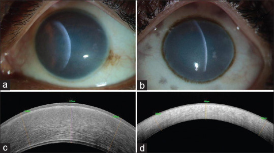
Congenital hereditary endothelial dystrophy (CHED) is a rare genetic corneal disorder causing progressive cornea clouding and significant visual impairment. CHED remains a leading indication for pediatric corneal transplantation despite its infrequency, particularly in regions with high consanguinity rates like Southeast Asia. Identifying the Solute Carrier Family 4 Member 11 (SLC4A11) gene as the genetic basis of CHED has led to the discovery of it's various genetic variations. However, a comprehensive understanding of its clinical-genetic correlation, pathophysiology, and optimal management is ongoing. This review aims to consolidate current knowledge about CHED, covering its genetic origins, pathophysiological mechanisms, clinical presentation, and management strategies. Surgical intervention, such as penetrating keratoplasty (PK), Descemet stripping automated endothelial keratoplasty (DSAEK), and Descemet membrane endothelial keratoplasty (DMEK), remains the primary treatment. DSAEK and DMEK offer advantages over PK, including quicker visual recovery, reduced complications, and longer graft survival, especially in the pediatric age group. The timing of surgical interventions depends on disease severity, age at presentation, comorbidities, and visual potential. Elevated oxidative stress in CHED corneal tissue suggests potential benefits from anti-inflammatory drugs to rescue mutated endothelial cells. Considering the limitations of corneal graft surgeries, exploring novel gene-based molecular therapies are essential for future management. Early diagnosis, appropriate surgical interventions, amblyopia control, and genetic counseling for predictive analysis are pivotal for optimizing CHED management. A multidisciplinary approach involving ophthalmologists, researchers, and genetic counselors is essential for precise diagnosis and optimal care for CHED patients.
Keywords: Congenital hereditary endothelial dystrophy; Solute Carrier Family 4 Member 11 (SLC4A11); corneal endothelial dystrophies in childhood.
Copyright: © 2023 Taiwan J Ophthalmol.
Conflict of interest statement
The authors declare that there are no conflicts of interests of this paper.
Figures


References
-
- Kenyon KR, Antine B. The pathogenesis of congenital hereditary endothelial dystrophy of the cornea. Am J Ophthalmol. 1971;72:787–95. - PubMed
-
- Maumenee AE. Congenital hereditary corneal dystrophy. Am J Ophthalmol. 1960;50:1114–24. - PubMed
-
- Antine B. Histology of congenital hereditary corneal dystrophy. Am J Ophthalmol. 1970;69:964–9. - PubMed
Publication types
LinkOut - more resources
Full Text Sources
Molecular Biology Databases