

Age influence on resistance and deformation of the human sutured meniscal horn in the immediate postoperative period
- PMID: 38249802
- PMCID: PMC10796521
- DOI: 10.3389/fbioe.2023.1249982
Age influence on resistance and deformation of the human sutured meniscal horn in the immediate postoperative period
Abstract
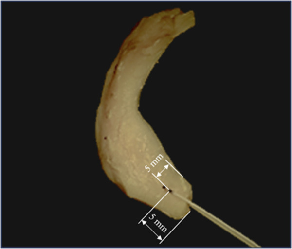
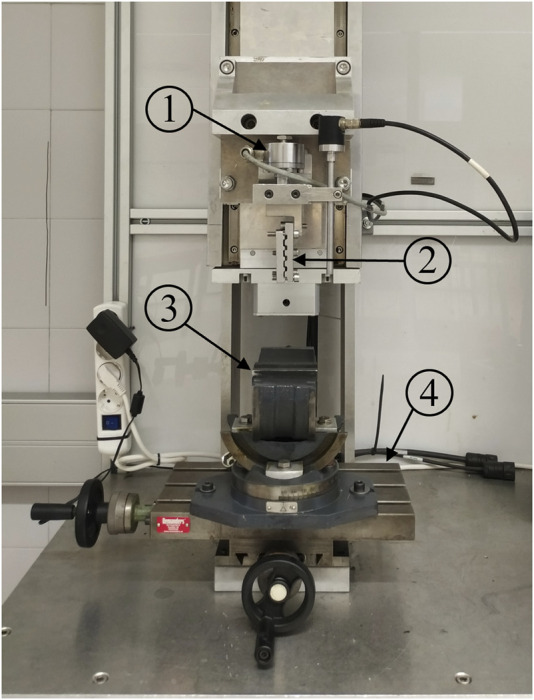
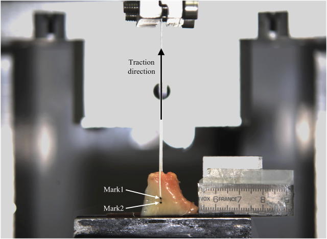
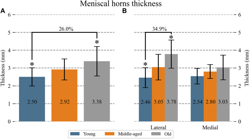
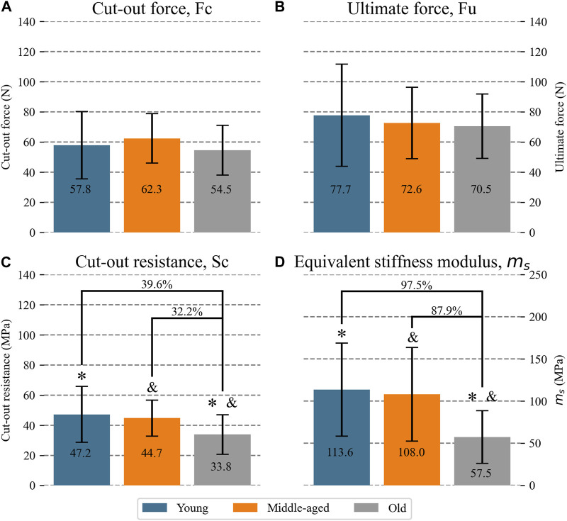
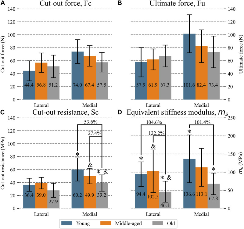
Introduction: To preserve knee function, surgical repair is indicated when a meniscal root disinsertion occurs. However, this surgery has not yet achieved complete recovery of the joint´s natural biomechanics, with the meniscus-suture interface identified as a potentially determining factor. Knowing the deformation and resistance behavior of the sutured meniscal horn and whether these properties are preserved as the patient ages could greatly contribute to improving repair outcomes. Methods: A cadaveric experimental study was conducted on human sutured menisci classified into three n = 22 age groups (young ≤55; 55 < middle-aged ≤75; 75 < old) were subjected to load-to-failure test by suture pulling. Meniscal thickness at the suture hole was measured and the applied traction force and tissue deformation in the suture area in the direction of traction were recorded during the test. The traction load that initiated the meniscal cut-out, , maximum load borne by the meniscus, , tissue stress at the cut-out initiation, , and equivalent stiffness modulus at the suture area, , were calculated. Results: At the tissue level, the resistance in terms of decrease with age (young: 47.2 MPa; middle-aged: 44.7 MPa; old: 33.8 MPa) being significantly different between the young and the old group (p = 0.015). Mean meniscal thickness increased with age (young: 2.50 mm; middle-aged: 2.92 mm; old: 3.38 mm; p = 0.001). Probably due to thickening, no differences in resistance were found at the specimen level, i.e., in (overall mean 58.2 N) and (overall mean 73.6 N). As for elasticity, was lower in the old group than in the young group (57.5 MPa vs. 113.6 MPa, p = 0.02) and the middle-aged one (57.5 MPa vs. 108.0 MPa, p = 0.04). Conclusion: Regarding the influence of age on the sutured meniscal horn tissue, in vitro experimentation revealed that meniscal horn specimens older than 75 years old had a more elastic tissue which was less resistant to cut-out than younger menisci at the suture hole area. However, a thickening of the meniscal horns with age, which was also found, leveled out the difference in the force that initiated the tear, as well as in the maximum force borne by the meniscus in the load-to-failure test.
Keywords: age influence; human meniscal tissue; meniscal root detachment; suture; tissue resistance.
Copyright © 2024 Peña-Trabalon, Perez-Blanca, Moreno-Vegas, Estebanez Campos and Prado-Novoa.
Conflict of interest statement
The authors declare that the research was conducted in the absence of any commercial or financial relationships that could be construed as a potential conflict of interest.
Figures
Similar articles
-
The Strength of Transosseous Medial Meniscal Root Repair Using a Simple Suture Technique Is Dependent on Suture Material and Position.Am J Sports Med. 2018 Mar;46(4):924-932. doi: 10.1177/0363546517749807. Epub 2018 Jan 24. Am J Sports Med. 2018. PMID: 29364699
-
Biomechanical Comparison of Two Different Sutures for the Tensile Strength of the Pullout Repair of Posterior Meniscal Root Tear.Cureus. 2023 Jul 24;15(7):e42378. doi: 10.7759/cureus.42378. eCollection 2023 Jul. Cureus. 2023. PMID: 37492035 Free PMC article.
-
Cyclic displacement after meniscal root repair fixation: a human biomechanical evaluation.Am J Sports Med. 2015 Apr;43(4):892-8. doi: 10.1177/0363546514562554. Epub 2015 Jan 2. Am J Sports Med. 2015. PMID: 25556220 Clinical Trial.
-
Arthroscopic meniscal repair with use of the outside-in technique.Instr Course Lect. 2000;49:195-206. Instr Course Lect. 2000. PMID: 10829175 Review.
-
Meniscal Repair Techniques.Clin Sports Med. 2020 Jan;39(1):37-56. doi: 10.1016/j.csm.2019.08.012. Clin Sports Med. 2020. PMID: 31767109 Review.
Cited by
-
Dynamic biomechanical effects of medial meniscus tears on the knee joint: a finite element analysis.J Orthop Surg Res. 2025 Jan 9;20(1):26. doi: 10.1186/s13018-024-05401-8. J Orthop Surg Res. 2025. PMID: 39780245 Free PMC article.
References
LinkOut - more resources
Full Text Sources
Research Materials