. 2023 Dec 30;15(12):6831-6847.
doi: 10.21037/jtd-23-1680. Epub 2023 Dec 26.

The molecular mechanism of sepsis-induced diaphragm dysfunction

Affiliations

The molecular mechanism of sepsis-induced diaphragm dysfunction

Xiaosa Yuan et al. J Thorac Dis. .

Abstract

Background: No effective drugs for the treatment of sepsis-induced diaphragm dysfunction are currently available. Therefore, it is particularly important to clarify the molecular regulatory mechanism of this condition and subsequently implement effective treatment and prevention of sepsis-induced diaphragm dysfunction.

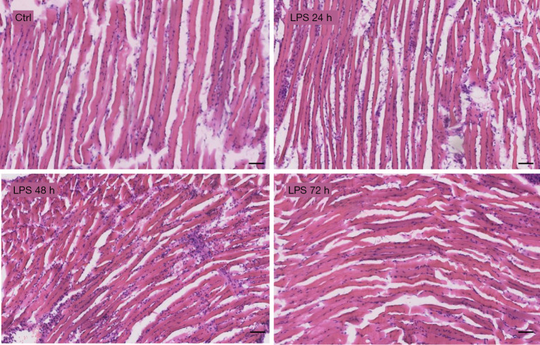
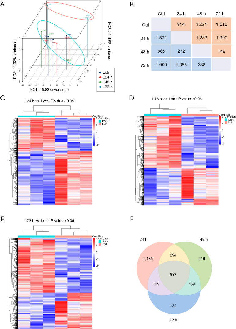
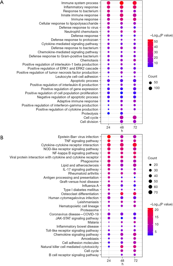
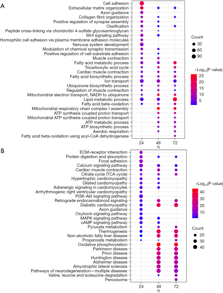
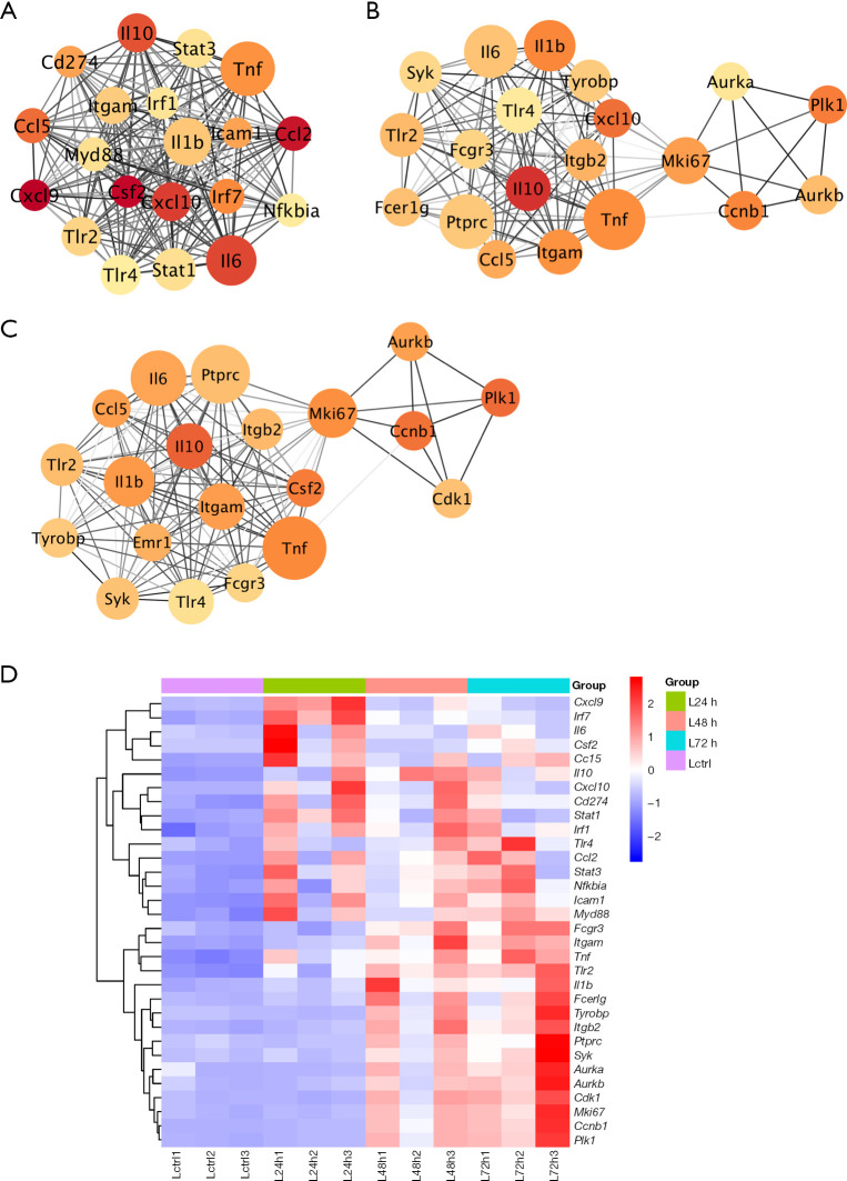
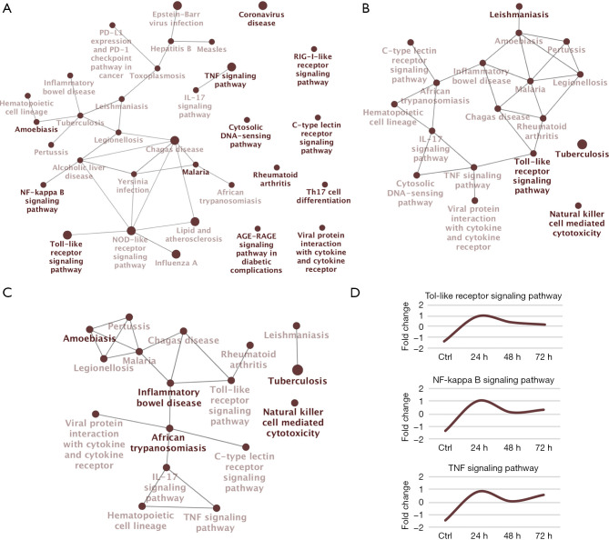
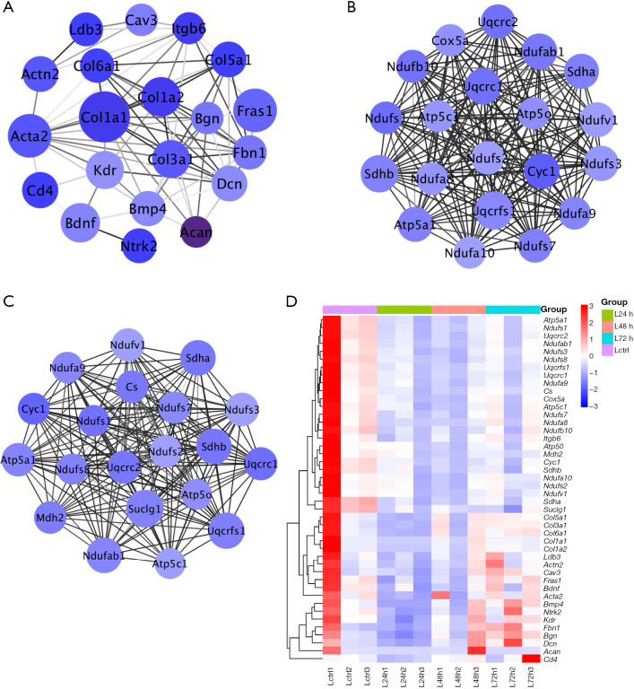
Methods: A mouse model of diaphragm dysfunction was established via injection of lipopolysaccharide (LPS). An RNA-sequencing (RNA-seq) technique was used to detect the differentially expressed genes (DEGs) in the diaphragms of mice. Gene Ontology (GO) and Kyoto Encyclopedia of Genes and Genomes (KEGG) enrichment analyses were performed for functional analysis of DEGs. The protein-protein interaction network obtained from the Search Tool for the Retrieval of Interacting Genes/Proteins (STRING) website was imported into Cytoscape, the key molecular regulatory network was constructed with CytoNCA, the ClueGo plugin was further used to analyze the core regulatory pathways of key molecular, and finally, the iRegulon plugin was used to the identify key transcription factors.

Results: The genes upregulated after LPS treatment were involved in biological processes and pathways related to immune response; the genes downregulated after LPS treatment were mainly correlated with the muscle contraction. The expressions of several inflammation-related genes were upregulated after LPS treatment, of which tumor necrosis factor (Tnf), interleukin (Il)-1β, and Il-6 assumed a core regulatory role in the network; meanwhile, the downregulated key genes included Col1a1, Uqcrfs1, Sdhb, and ATP5a1, among others. These key regulatory factors participated in the activation of Toll-like receptor (TLR) signaling pathway, nuclear factor (NF)-κB signaling pathway, and TNF signaling pathway as well as the inhibition of oxidative phosphorylation pathway, cardiac muscle contraction pathway, and citrate cycle pathway. Finally, RelA, IRF1, and STAT3, were identified as the key regulators in the early stage of diaphragmatic inflammatory response.

Conclusions: Sepsis-induced diaphragm dysfunction in mice is closely correlated with the activation of TLR signaling pathway, NF-κB signaling pathway, and TNF signaling pathway and the inhibition of oxidative phosphorylation pathway, cardiac muscle contraction pathway, and citrate cycle pathway. Our findings provide insight into the molecular mechanism of sepsis-induced diaphragm dysfunction in mice and provide a promising new strategy for targeted treatment of diaphragm dysfunction.

Keywords: Diaphragm dysfunction; energy metabolism; inflammation; sepsis.


Conflict of interest statement

Conflicts of Interest: All authors have completed the ICMJE uniform disclosure form (available at https://jtd.amegroups.com/article/view/10.21037/jtd-23-1680/coif). The authors have no conflicts of interest to declare.

Figures

Figure 1
Figure 1
Changes in diaphragmatic structure after LPS treatment. Bar =50 μm. 12-μm thick frozen sections of diaphragm tissue were stained with HE. Ctrl, control; LPS, lipopolysaccharide; HE, hematoxylin and eosin.
Figure 2
Figure 2
Analysis of DEGs in the mouse diaphragm after LPS treatment. (A) PCA analysis. (B) Comparison of the number of DEGs in the mouse diaphragm of the LPS-treatment and control group at different time points after treatment. (C-E) Differential gene expression heatmaps at different time points after LPS treatment. (F) Venn diagram analysis of the number of differentially co-expressed genes at different time points after LPS treatment. PC, principal component; ctrl, control; DEG, differentially expressed gene; LPS, lipopolysaccharide; PCA, principal component analysis.
Figure 3
Figure 3
Functional analysis of DEGs in the diaphragm upregulated after LPS treatment. (A) GO biological process enriched by the genes upregulated at different time points. (B) KEGG enrichment analysis of genes upregulated at different time points. The circle’s size and color indicate the number and −log10(P value) of DEGs annotated in the specific terms, respectively. ERK, extraneous signal-regulated kinase; TNF, tumor necrosis factor; NOD, nucleotide oligomerization domain; NF, nuclear factor; IL, interleukin; COVID-19, coronavirus disease 2019; JAK-STAT, Janus kinase-signal transducer and activator of transcription; DEG, differentially expressed gene; LPS, lipopolysaccharide; GO, Gene Ontology; KEGG, Kyoto Encyclopedia of Genes and Genomes.
Figure 4
Figure 4
Functional analysis of the DEGs in the diaphragm downregulated after LPS treatment. (A) The GO biological processes enriched by the genes downregulated at different time points. (B) KEGG enrichment analysis of genes downregulated at different time points. The circle’s size and color indicate the number and −log10(P value) of DEGs annotated in the specific terms, respectively. NADH, reduced forms of the nicotinamide adenine dinucleotide; ATP, adenosine triphosphate; CoA, coenzyme A; ECM, extracellular matrix; TCA, tricarboxylic acid; MAPK, mitogen-activated protein kinase; cAMP, cyclic adenosine monophosphate; DEG, differentially expressed gene; LPS, lipopolysaccharide; GO, Gene Ontology; KEGG, Kyoto Encyclopedia of Genes and Genomes.
Figure 5
Figure 5
Analysis of key genes upregulated in the diaphragm at different time points after LPS treatment. (A) Analysis of key genes in the diaphragm at 24 h after LPS treatment. (B) Analysis of key genes in the diaphragm at 48 h after LPS treatment. (C) Analysis of key genes in the diaphragm at 72 h after LPS treatment. (D) A heat map of the expression levels of key genes upregulated in diaphragm at different time points after LPS treatment. The circle’s size and color indicate the degree and fold of DEGs, respectively. Ctrl, control; LPS, lipopolysaccharide; DEG, differentially expressed gene.
Figure 6
Figure 6
Analysis of key pathways upregulated in the diaphragm at different time points after LPS treatment. (A) Analysis of key pathways upregulated in the diaphragm at 24 h after LPS treatment. (B) Analysis of key pathways upregulated in the diaphragm at 48 h after LPS treatment. (C) Analysis of key pathways upregulated in diaphragm at 72 h after LPS treatment. (D) The average expression profiles of DEGs involved in the major KEGG pathways. The circle’s size indicates the number of DEGs annotated in the given terms. PD-L1, programmed cell death ligand 1; PD-1, programmed cell death protein 1; TNF, tumor necrosis factor; IL, interleukin; NF, nuclear factor; NOD, nucleotide oligomerization domain; AGE, advanced glycation end products; RAGE, receptor for advanced glycation end products; ctrl, control; LPS, lipopolysaccharide; DEG, differentially expressed gene; KEGG, Kyoto Encyclopedia of Genes and Genomes.
Figure 7
Figure 7
Analysis of key genes downregulated in diaphragm at different time points after LPS treatment. (A) Analysis of key genes downregulated in the diaphragm at 24 h after LPS treatment. (B) Analysis of key genes downregulated in diaphragm at 48 h after LPS treatment. (C) Analysis of key genes downregulated in diaphragm at 72 h after LPS treatment. (D) A heat map of key genes downregulated in the diaphragm at different time points after LPS treatment. The circle’s size and color indicate the degree and fold change of DEGs, respectively. Ctrl, control; LPS, lipopolysaccharide; DEG, differentially expressed gene.
Figure 8
Figure 8
Analysis of the key pathways downregulated in the diaphragm at different time points after LPS treatment. (A) Analysis of key pathways downregulated in the diaphragm at 24 h after LPS treatment. (B) Analysis of key pathways downregulated in the diaphragm at 48 h after LPS treatment. (C) Analysis of key pathways downregulated in the diaphragm at 72 h after LPS treatment. (D) The average expression profiles of DEGs involved in major KEGG pathways. The circle’s size indicates the number of DEGs annotated in the given terms. ECM, extracellular matrix; TCA, tricarboxylic acid; ctrl, control; LPS, lipopolysaccharide; DEG, differentially expressed gene; KEGG, Kyoto Encyclopedia of Genes and Genomes.
Figure 9
Figure 9
Analysis of key transcription factors at 24 h after LPS treatment. (A) Network of the key molecules regulated by RelA. (B) Network of the key molecules regulated by Irf1. (C) Network of the key molecules regulated by Stat3. LPS, lipopolysaccharide.

Similar articles

Cited by

References

    1. Oliveira TS, Santos AT, Andrade CBV, et al. Sepsis Disrupts Mitochondrial Function and Diaphragm Morphology. Front Physiol 2021;12:704044. 10.3389/fphys.2021.704044 - DOI - PMC - PubMed
    1. Lelubre C, Vincent JL. Mechanisms and treatment of organ failure in sepsis. Nat Rev Nephrol 2018;14:417-27. 10.1038/s41581-018-0005-7 - DOI - PubMed
    1. Baldwin CE, Bersten AD. Alterations in respiratory and limb muscle strength and size in patients with sepsis who are mechanically ventilated. Phys Ther 2014;94:68-82. 10.2522/ptj.20130048 - DOI - PubMed
    1. Wang W, Shen D, Zhang L, et al. SKP-SC-EVs Mitigate Denervated Muscle Atrophy by Inhibiting Oxidative Stress and Inflammation and Improving Microcirculation. Antioxidants (Basel) 2021;11:66. 10.3390/antiox11010066 - DOI - PMC - PubMed
    1. Petrof BJ. Diaphragm Weakness in the Critically Ill: Basic Mechanisms Reveal Therapeutic Opportunities. Chest 2018;154:1395-403. 10.1016/j.chest.2018.08.1028 - DOI - PubMed