. 2024 Jan 1;14(3):1049-1064.
doi: 10.7150/thno.89180. eCollection 2024.

Keratinocyte-to-macrophage communication exacerbate psoriasiform dermatitis via LRG1-enriched extracellular vesicles

Affiliations

Keratinocyte-to-macrophage communication exacerbate psoriasiform dermatitis via LRG1-enriched extracellular vesicles

Wenjuan Jiang et al. Theranostics. .

Abstract

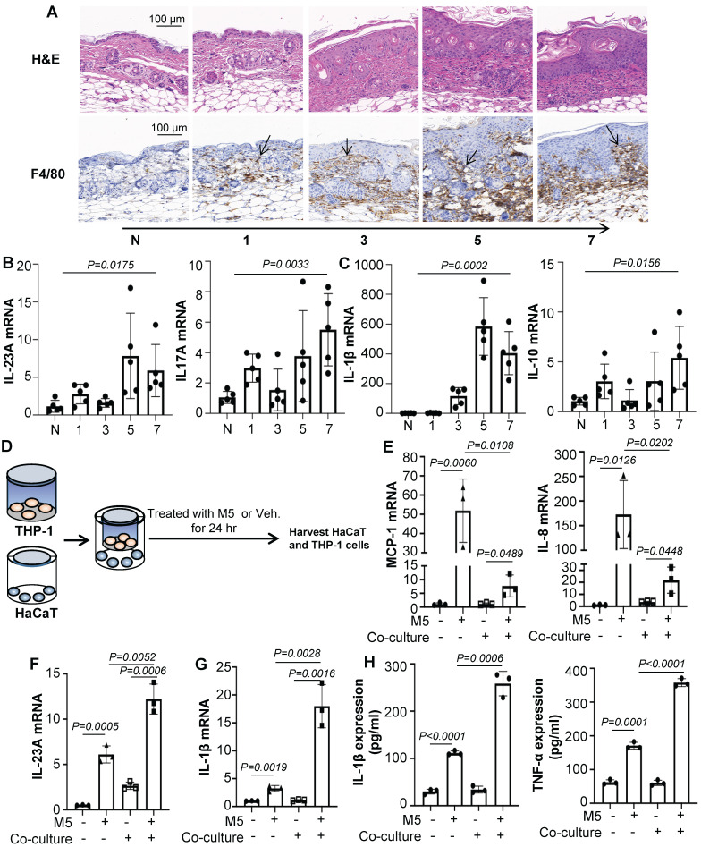
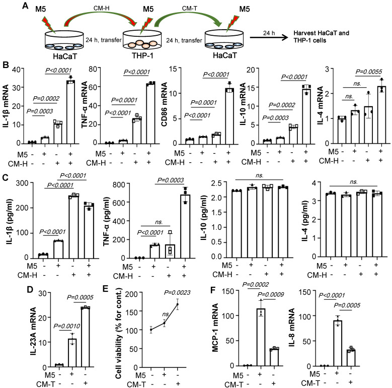
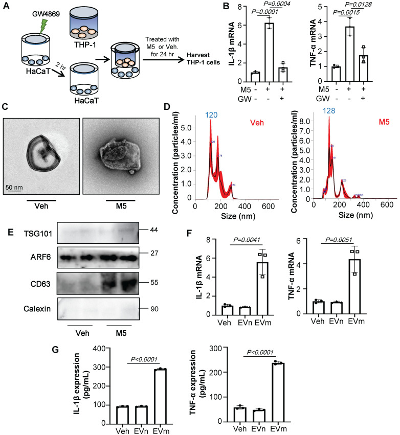
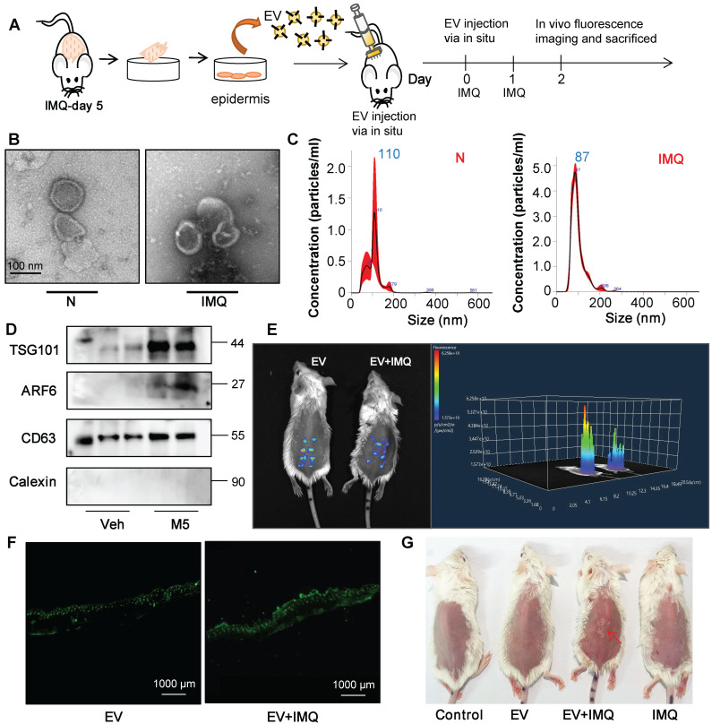
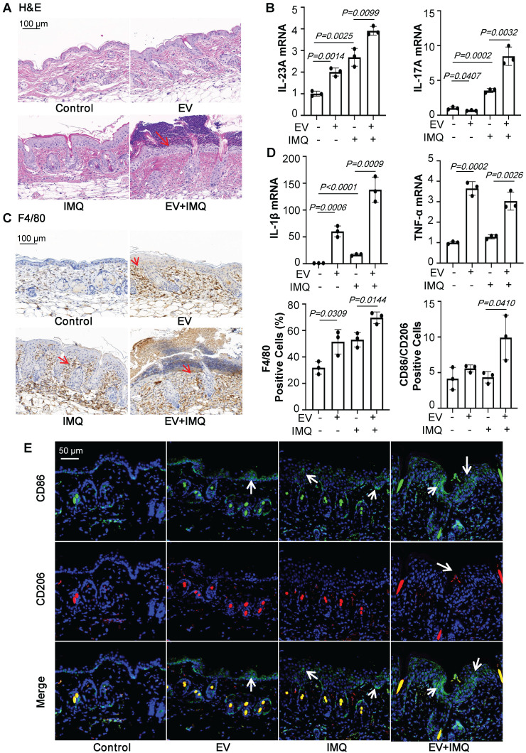
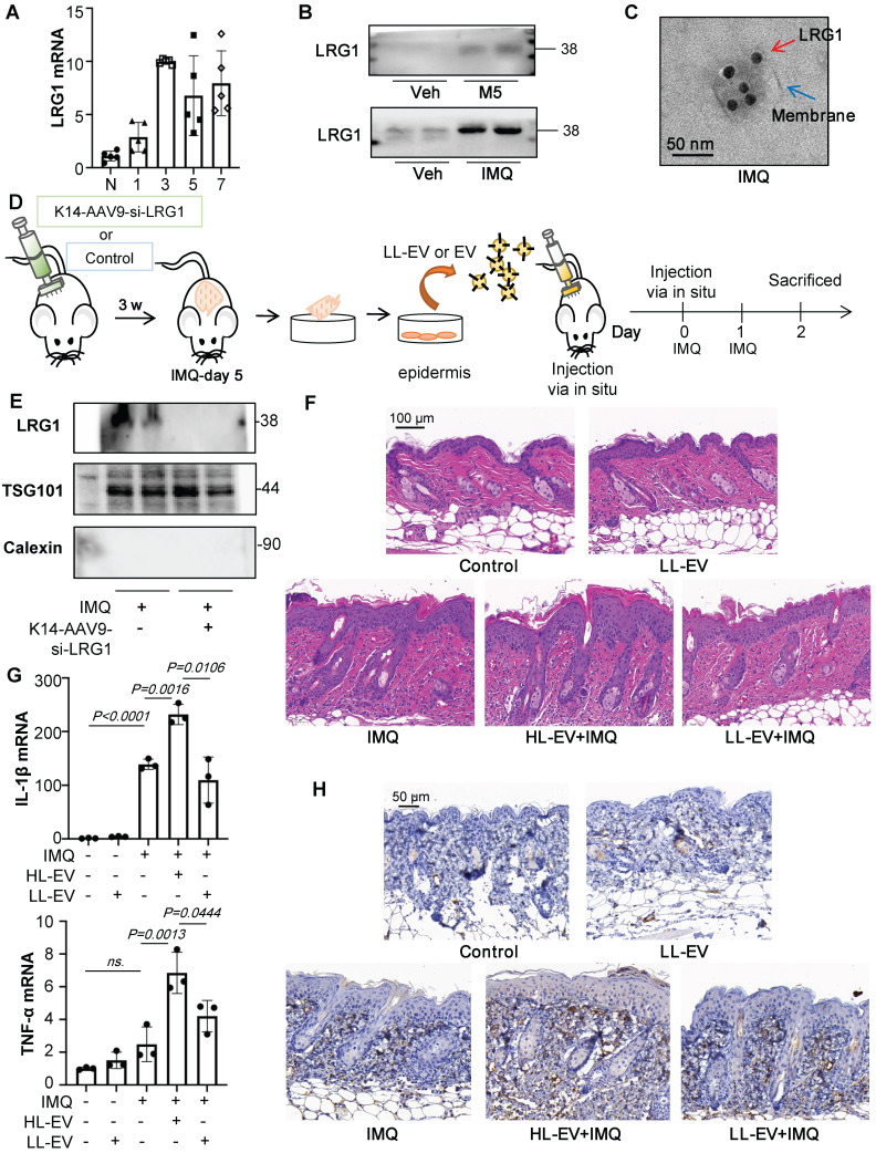
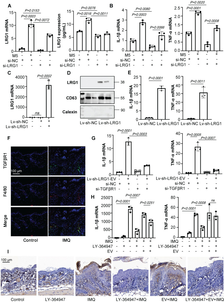
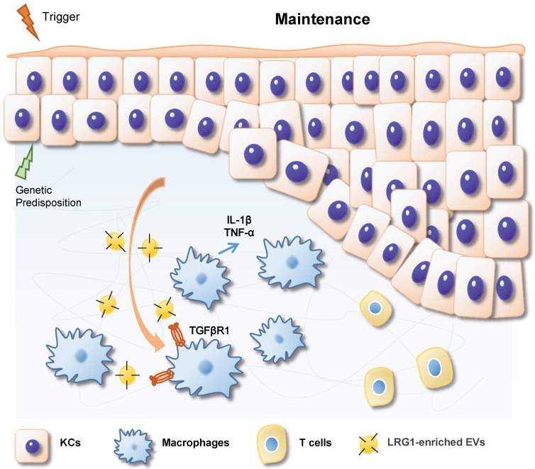
Rationale: Macrophage-associated inflammation and keratinocytes excessive proliferation and inflammatory cytokines secretion induced by stimulation play an important role in the progression of psoriasiform dermatitis. However, how these two types of cells communicate remains obscure. Methods: We induced a mouse model with experimental psoriasiform dermatitis by Imiquimod (IMQ). To investigate whether damaged keratinocytes promote macrophage polarization and accelerate skin lesions by releasing extracellular vesicle (EV), purified EV were isolated from the primary epidermis of 5-day IMQ-induced psoriasiform dermatitis model mice, and then fluorescence-labeled the EV with PKH67. The EV was injected into the skin of mice treated with IMQ or vehicle 2 days in situ. In addition, we established a co-culture system of the human monocytic cell line (THP-1) and HaCaT, and THP-1/HaCaT conditioned media culture model in vitro respectively. Subsequently, we evaluated the effect of Leucine-rich α-2-glycoprotein 1 (LRG1)-enriched EV on macrophage activation. Results: We demonstrated macrophages can significantly promote keratinocyte inflammation and macrophage polarization may be mediated by intercellular communication with keratinocytes. Interestingly, IMQ-induced 5-day, keratinocyte-derived EV recruited macrophage and enhanced the progression of skin lesions. Similar to results in vivo, EV released from M5-treated HaCaT significantly promotes Interleukin 1β (IL-1β) and Tumor necrosis factor α (TNF-α) expression of THP-1 cells. Importantly, we found that LRG1-enriched EV regulates macrophages via TGF beta Receptor 1 (TGFβR1) dependent process. Conclusion: Our findings indicated a novel mechanism for promoting psoriasiform dermatitis, which could be a potential therapeutic target.

Keywords: Extracellular vesicle; Keratinocyte.; Leucine-rich α-2-glycoprotein 1; Macrophage; Psoriasis.


Conflict of interest statement

Competing Interests: The authors have declared that no competing interest exists.

Figures

Figure 1
Figure 1
Macrophages participate in psoriasiform dermatitis. (A) Mice skin tissues stained with H&E and F4/80. Scale bar, 100 μm. (B) IL-23A, IL-17A mRNA was assessed by quantitative real-time PCR. (C) IL-1β, IL-10 mRNA was assessed by quantitative real-time PCR. (D) HaCaT and THP-1 co-culture system. THP-1 cells were differentiated using PMA pre-treatment for 24 h. (E) The level of MCP-1, IL-8 in HaCaT cells analysed by quantitative real-time PCR. (F) The level of IL-23A in HaCaT cells was analysed by quantitative real-time PCR. (G) The level of IL-1β in THP-1 cells was analysed by quantitative real-time PCR. (H) The protein levels of IL-1β and TNF-α in the co-culture supernatant were detected by ELISA. Similar results were obtained in 3 independent experiments with 5 mice per group or in triplicate culture assays.
Figure 2
Figure 2
M5-stimulated Keratinocytes induce macrophage polarization. (A) HaCaT/THP-1 conditioned media culture model. THP-1 cells were differentiated by PMA pre-treatment for 24 h. (B) The mRNA levels of IL-1β, TNF-α, CD86, IL-10, IL-4 in THP-1 cells analysed by quantitative real-time PCR. (C) The protein levels of IL-1β, TNF-α, IL-10, IL-4 in THP-1 cell supernatant were detected by ELISA. (D) The mRNA level of IL-23A in HaCaT cells analysed by quantitative real-time PCR. (E) Cell viability detected by CCK-8 kit. (F) The mRNA levels of MCP-1, IL-8 in HaCaT cells analysed by quantitative real-time PCR. Similar results were obtained in 3 independent experiments or in triplicate culture assays.
Figure 3
Figure 3
Keratinocyte promotes macrophage polarization by secrete EVs. (A) GW4869 pre-treated HaCaT and THP-1 co-culture system. THP-1 cells were differentiated using PMA pre-treatment for 24 h. (B) The level of IL-1β, TNF-α in THP-1 cells analysed by quantitative real-time PCR. (C) Transmission electron photomicrographs of HaCaT-derived EV. (D) HaCaT-derived EV representative image with nanoparticle tracking analysis (NTA). (E) Expression of TSG101, ARF6, CD63 and Calexin in HaCaT-derived EV were detected by western blot. (F) The level of IL-1β, TNF-α in THP-1 cells treated with HaCaT-derived EV analysed by quantitative real-time PCR. (G) The protein levels of IL-1β and TNF-α in THP-1 cell supernatant were detected by ELISA. Similar results were obtained in 3 independent experiments or in triplicate culture assays.
Figure 4
Figure 4
Keratinocyte-derived EVs promote IMQ-induced psoriasiform dermatitis. (A) Purified EVs were isolated from primary keratinocyte of 5-day IMQ-induced mice and PKH67-labelled EVs were injected into mice via skin in situ injection. (B) Transmission electron photomicrographs of primary keratinocytes-derived EV of IMQ-induced mice. (C) Primary keratinocytes-derived EV of IMQ-induced mice representative image with nanoparticle tracking analysis (NTA). (D) Expression of TSG101, ARF6, CD63 and Calexin in primary keratinocytes-derived EV of IMQ-induced mice were detected by western blot. (E) Imaging of PKH67-labeled EVs in mice. About 100 μg (at protein level) in 100 μL EV from IMQ exposured mice primary keratinocytes, labeled with PKH67 were injected via in situ. About 48 h after the in situ injection, in vivo fluorescence imaging were performed. (F) Fluorescence imaging were performed in skin tissue section. Scale bar, 1000 μm. (G) Photos of skin lesions on the back of mice. Similar results were obtained in 3 independent experiments with 3 mice per group or in triplicate culture assays.
Figure 5
Figure 5
Keratinocyte-derived EVs promote M1 macrophage polarization and psoriasiform dermatitis. (A) Mice skin tissues stained with H&E. Scale bar, 100 μm. (B) IL-23A, IL-17A mRNA was assessed by quantitative real-time PCR. (C) Mice skin tissues stained with F4/80. Scale bar, 100 μm. (D) IL-1β, TNF-α mRNA was assessed by quantitative real-time PCR. (E) Mice skin tissues stained with CD86 and CD206. Scale bar, 50 μm. Similar results were obtained in 3 independent experiments with 3 mice per group.
Figure 6
Figure 6
LRG1-enriched EVs promote M1 macrophage polarization and psoriasiform dermatitis. (A) The level of LRG1 in IMQ-induced mouse model analysed by quantitative real-time PCR. (B) Expression of LRG1 in HaCaT-derived EV and primary keratinocytes-derived EV were detected by western blot. (C) Representative transmission electron photomicrographs immunogold- labeled with an anti-LRG1 antibody of primary keratinocytes-derived EV. Scale bar: 50 nm. (D) The AAV9 vectors delivering si-LRG1 driven by Keratin 14 (K14) promoter induce to knockdown the expression of LRG1 of IMQ-treated mice by skin in situ injection, released EVs were isolated from primary epidermis cells culture. (E) Expression of TSG101, LRG1 and Calexin in primary keratinocytes-derived EV of IMQ-induced mice were detected by western blot. (F) Mice skin tissues stained with H&E. Scale bar, 100 μm. (G) IL-1β, TNF-α mRNA was assessed by quantitative real-time qPCR. (H) Mice skin tissues stained with F4/80. Scale bar, 50 μm. Similar results were obtained in 3 independent experiments with 3 mice per group.
Figure 7
Figure 7
LRG1-enriched EVs from Keratinocytes mediate macrophage polarization via TGFβR1. (A) The level of LRG1 in HaCaT cells treated with si-LRG1 analysed by quantitative real-time PCR and ELISA. (B) The mRNA levels of IL-1β, TNF-α in THP-1 cells treated with conditional medium from si-LRG1-stimulated HaCaT cells analysed by quantitative real-time PCR. (C) The level of LRG1 in HaCaT cells treated with Lv-sh-LRG1 analysed by quantitative real-time PCR. (D) The level of LRG1 in EV derived from HaCaT treated with Lv-sh-LRG1 detected by western blot. (E) The mRNA levels of IL-1β, TNF-α in THP-1 cells treated with EV derived from Lv-sh-LRG1-stimulated HaCaT cells analysed by quantitative real-time PCR. (F) Mice skin tissues stained with TGFβR1. Scale bar, 100 μm. (G) The mRNA levels of IL-1β, TNF-α in si-TGFβR1 pre-treated THP-1 cells treated with EV derived from Lv-sh-LRG1-stimulated HaCaT cells analysed by quantitative real-time PCR. (H) IL-1β, TNF-α mRNA was assessed by quantitative real-time PCR. (I) Mice skin tissues stained with F4/80. Scale bar, 100 μm. Similar results were obtained in 3 independent experiments with 3 mice per group or in triplicate culture assays.
Figure 8
Figure 8
During IMQ-induced psoriasiform dermatitis progression, LRG1-enriched EVs released from keratinocytes regulates macrophage by a TGFβR1-dependent pathway and subsequently up-regulates expression of some inflammatory genes, thereby promoting psoriasiform dermatitis.

Similar articles

Cited by

References

    1. Griffiths CEM, Armstrong AW, Gudjonsson JE, Barker J. Psoriasis. Lancet. 2021;397:1301–15. - PubMed
    1. Boehncke WH, Schon MP. Psoriasis. Lancet. 2015;386:983–94. - PubMed
    1. Ni X, Lai Y. Keratinocyte: A trigger or an executor of psoriasis? J Leukoc Biol. 2020;108:485–91. - PubMed
    1. Leite Dantas R, Masemann D, Schied T, Bergmeier V, Vogl T, Loser K. et al. Macrophage-mediated psoriasis can be suppressed by regulatory T lymphocytes. J Pathol. 2016;240:366–77. - PubMed
    1. Kuraitis D, Rosenthal N, Boh E, McBurney E. Macrophages in dermatology: pathogenic roles and targeted therapeutics. Arch Dermatol Res. 2022;314:133–40. - PubMed