

Observations on the transmission of Dientamoeba fragilis and the cyst life cycle stage
- PMID: 38250789
- PMCID: PMC11007279
- DOI: 10.1017/S0031182024000076
Observations on the transmission of Dientamoeba fragilis and the cyst life cycle stage
Abstract
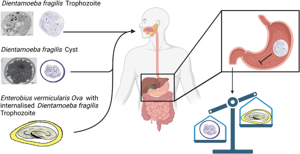
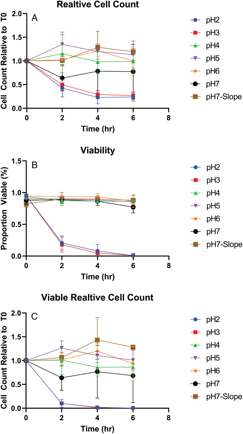
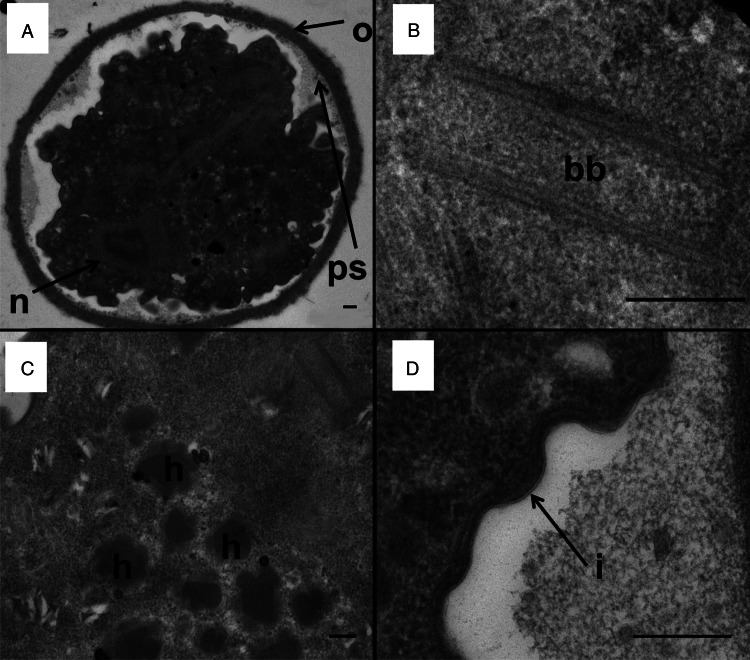
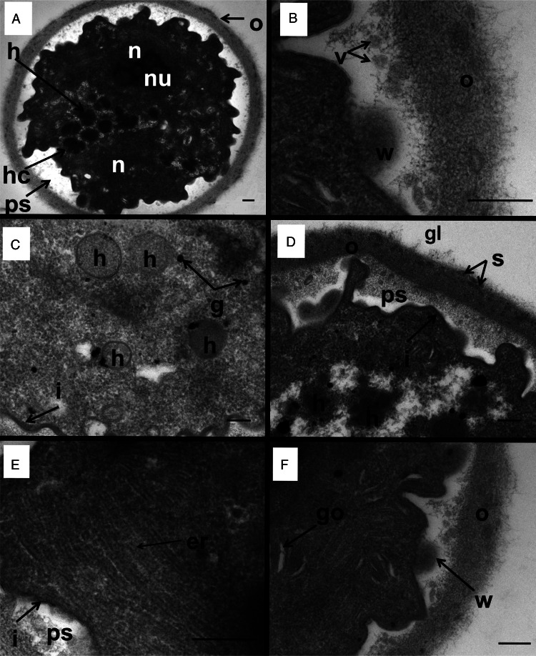
Little is known about the life cycle and mode of transmission of Dientamoeba fragilis. Recently it was suggested that fecal–oral transmission of cysts may play a role in the transmission of D. fragilis. In order to establish an infection, D. fragilis is required to remain viable when exposed to the pH of the stomach. In this study, we investigated the ability of cultured trophozoites to withstand the extremes of pH. We provide evidence that trophozoites of D. fragilis are vulnerable to highly acidic conditions. We also investigated further the ultrastructure of D. fragilis cysts obtained from mice and rats by transmission electron microscopy. These studies of cysts showed a clear cyst wall surrounding an encysted parasite. The cyst wall was double layered with an outer fibrillar layer and an inner layer enclosing the parasite. Hydrogenosomes, endoplasmic reticulum and nuclei were present in the cysts. Pelta-axostyle structures, costa and axonemes were identifiable and internal flagellar axonemes were present. This study therefore provides additional novel details and knowledge of the ultrastructure of the cyst stage of D. fragilis.
Keywords: Dientamoeba fragilis; cyst; electron microscopy; transmission.
Conflict of interest statement
None.
Figures
References
-
- Abd H (2021) Dientamoeba fragilis trophozoites undergoing encystation, apoptosis and necrosis as human parasitic amoeba in clinical stool samples. European Academic Research IX, 4633–4649.
-
- Banik GR, Birch D, Stark D and Ellis JT (2012) A microscopic description and ultrastructural characterisation of Dientamoeba fragilis: an emerging cause of human enteric disease. International Journal for Parasitology 42, 139–153. - PubMed
-
- Barratt JLN, Banik GR, Harkness J, Marriott D, Ellis JT and Stark D (2010) Newly defined conditions for the in vitro cultivation and cryopreservation of Dientamoeba fragilis: new techniques set to fast track molecular studies on this organism. Parasitology 137, 1867–1878. - PubMed
-
- Barratt JL, Harkness J, Marriott D, Ellis JT and Stark D (2011) The ambiguous life of Dientamoeba fragilis: the need to investigate current hypotheses on transmission [review]. Parasitology 138, 557–572. - PubMed
-
- Barratt JL, Cao M, Stark DJ and Ellis JT (2015) The transcriptome sequence of Dientamoeba fragilis offers new biological insights on its metabolism, kinome, degradome and potential mechanisms of pathogenicity. Protist 166, 389–408. - PubMed
MeSH terms
LinkOut - more resources
Full Text Sources
