Review
. 2024 Jan 18;16(1):55.
doi: 10.3390/toxins16010055.

Voltage-Gated Sodium Channel Inhibition by µ-Conotoxins

Affiliations
Review

Voltage-Gated Sodium Channel Inhibition by µ-Conotoxins

Kirsten L McMahon et al. Toxins (Basel). .

Abstract

µ-Conotoxins are small, potent pore-blocker inhibitors of voltage-gated sodium (NaV) channels, which have been identified as pharmacological probes and putative leads for analgesic development. A limiting factor in their therapeutic development has been their promiscuity for different NaV channel subtypes, which can lead to undesirable side-effects. This review will focus on four areas of µ-conotoxin research: (1) mapping the interactions of µ-conotoxins with different NaV channel subtypes, (2) µ-conotoxin structure-activity relationship studies, (3) observed species selectivity of µ-conotoxins and (4) the effects of µ-conotoxin disulfide connectivity on activity. Our aim is to provide a clear overview of the current status of µ-conotoxin research.

Keywords: disulfide-rich peptide; peptide; structure–activity relationships; subtype selectivity; venom peptide; voltage-gated sodium channels; µ-conotoxin.


Conflict of interest statement

C.I.S. is an employee of Genentech Inc. and a shareholder of Roche. K.L.M. and I.V. declare no conflict of interest.

Figures
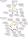
Figure 3
Figure 3
Summary of µ-conotoxin residues influencing potency and selectivity, as determined by structure–activity relationship studies. Representative globular fold of M4-branch (top panel) and M5-branch (bottom panel) µ-conotoxins, as modelled by available NMR structures. Conserved residues (>90%) are represented by single amino acid letter (navy circles) or + (either Arg or Lys; purple circles). Non-conserved residues are not annotated (teal circles). Cysteine residues (yellow circles) and disulfide bonds (yellow dashed line) are represented with canonical connectivity. Structure–activity relationship studies have identified several residues that modulate inhibition (pink dashed rings). Summary of the key findings are shown in neighbouring boxes. Hyp—hydroxyproline, Pyr—pyroglutamate and Sar—N-methylglycine. For clarity, disulfide bond mutations, which will be discussed in the following section, are not included. (Hui, 2002 [18]; Shon, 1998 [28]; Lewis, 2007 [30]; Schroeder, 2008 [40]; Harvey, 2018 [41]; McMahon, 2022 [44]; Sato, 1991 [48]; Schroeder, 2012 [65]; Zhang, 2007 [66]; MaArthur, 2011 [67]; Sato, 2014 [68]; Van der Haegan, 2011 [69]; Nakamura, 2001 [71]; Walewska, 2013 [72]; Yao, 2008 [77]; Akondi, 2014 [80]; Kuang, 2013 [81]; Green, 2014 [82]; Han, 2009 [83]).
Figure 1
Figure 1
Distribution of human NaV channels and associated channelopathies. The nine NaV channel subtypes (NaV1.1–1.9; teal segments) are encoded by a corresponding SCNxA gene (where x = 1–5, 8–11). Major tissues expressing various NaV subtypes are indicated by dotted lines. Outer segments denote channelopathies associated with NaV channel mutations (pain—dark blue, cardiac—purple, epilepsy—orange, and skeletal muscle disorders—yellow). DRG—dorsal root ganglia.
Figure 2
Figure 2
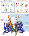
Representations of µ-conotoxin 3D NMR structures determined to date (top) and illustrative µ-conotoxin–NaV channel binding site (bottom). Coordinates from the PDB (1TCJ GIIIA; 1GIB GIIIB; 6MJD GIIIC; 1R9I PIIIA; 2LOC BuIIIB, 2YEN CnIIIC; 2LXG KIIIA; 1Q2J SmIIIA, 6X8R SxIIIC and 6J8J hNaV1.7) and BMRB (20024 TIIIA and 20025 SIIIA) were used to generate figures in PyMol.
Figure 4
Figure 4
Sequence differences within the µ-conotoxin NaV channel binding site may confer species specificity. Only minimal sequence differences are observed between mammalian species within the NaV channel binding site. For clarity, only the human, rat, and mouse sequences for three subtypes are shown, including the frequently studied NaV1.2 and NaV1.4, as well as the therapeutically relevant NaV1.7. Sequence differences are highlighted by shaded bars. Residues are coloured according to properties (positively charged—blue; negatively charged—red and neutral—white).

References

    1. Puillandre N., Duda T.F., Meyer C., Olivera B.M., Bouchet P. One, four or 100 genera? A new classification of the cone snails. J. Molluscan Stud. 2015;81:1–23. doi: 10.1093/mollus/eyu055. - DOI - PMC - PubMed
    1. Olivera B.M., Gray W.R., Zeikus R., McIntosh J.M., Varga J., Rivier J., de Santos V., Cruz L.J. Peptide neurotoxins from fish-hunting cone snails. Science. 1985;230:1338–1343. doi: 10.1126/science.4071055. - DOI - PubMed
    1. Olivera B.M., Seger J., Horvath M.P., Fedosov A.E. Prey-capture strategies of fish-hunting cone snails: Behavior, neurobiology and evolution. Brain Behav. Evol. 2015;86:58–74. doi: 10.1159/000438449. - DOI - PMC - PubMed
    1. Puillandre N., Koua D., Favreau P., Olivera B.M., Stocklin R. Molecular phylogeny, classification and evolution of conopeptides. J. Mol. Evol. 2012;74:297–309. doi: 10.1007/s00239-012-9507-2. - DOI - PubMed
    1. Safavi-Hemami H., Brogan S.E., Olivera B.M. Pain therapeutics from cone snail venoms: From Ziconotide to novel non-opioid pathways. J. Proteom. 2019;190:12–20. doi: 10.1016/j.jprot.2018.05.009. - DOI - PMC - PubMed

MeSH terms

LinkOut - more resources