. 2024 Jan 22;81(1):49.
doi: 10.1007/s00018-023-05067-1.

Selenium-SelK-GPX4 axis protects nucleus pulposus cells against mechanical overloading-induced ferroptosis and attenuates senescence of intervertebral disc

Affiliations

Selenium-SelK-GPX4 axis protects nucleus pulposus cells against mechanical overloading-induced ferroptosis and attenuates senescence of intervertebral disc

Chunwang Jia et al. Cell Mol Life Sci. .

Abstract

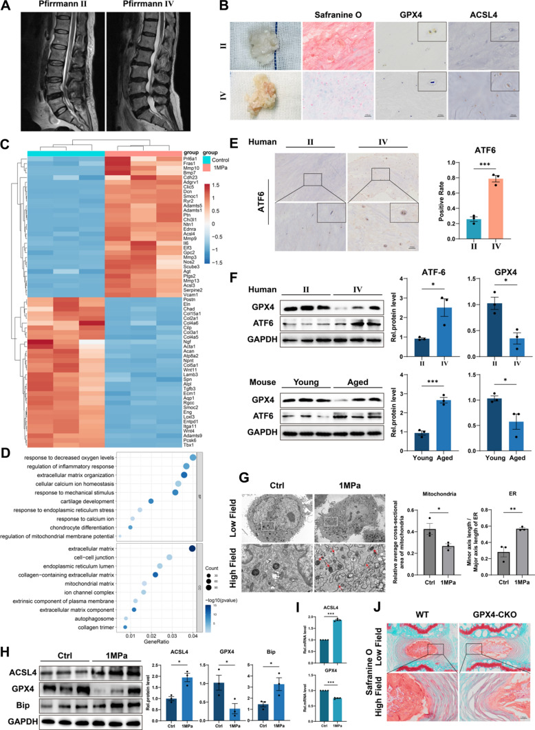
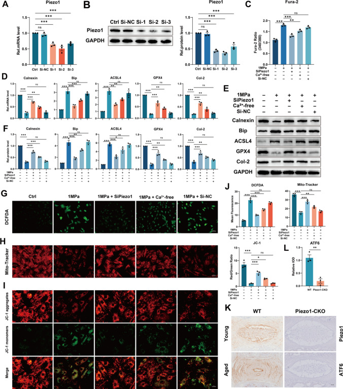
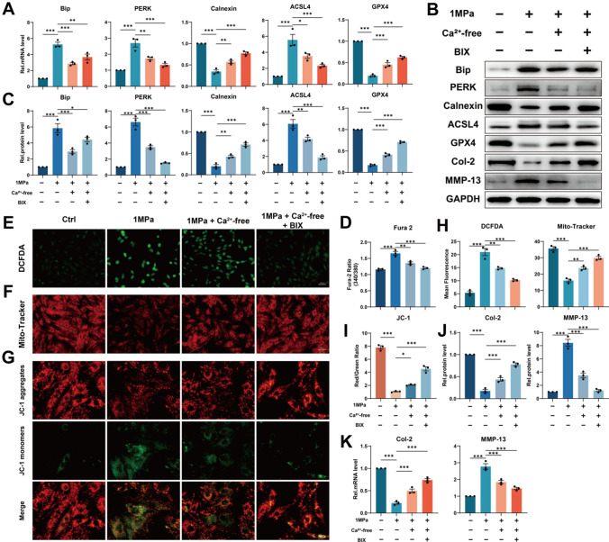
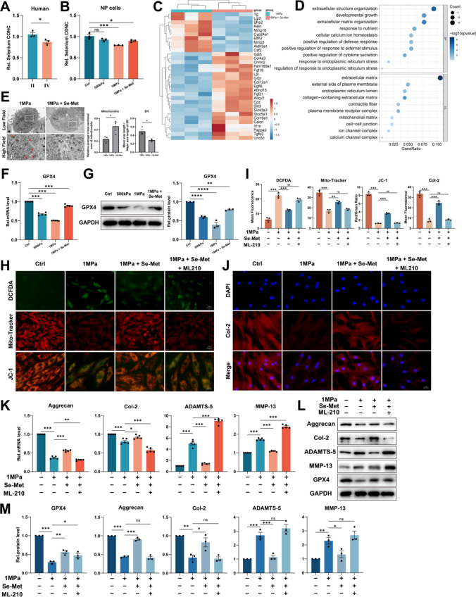
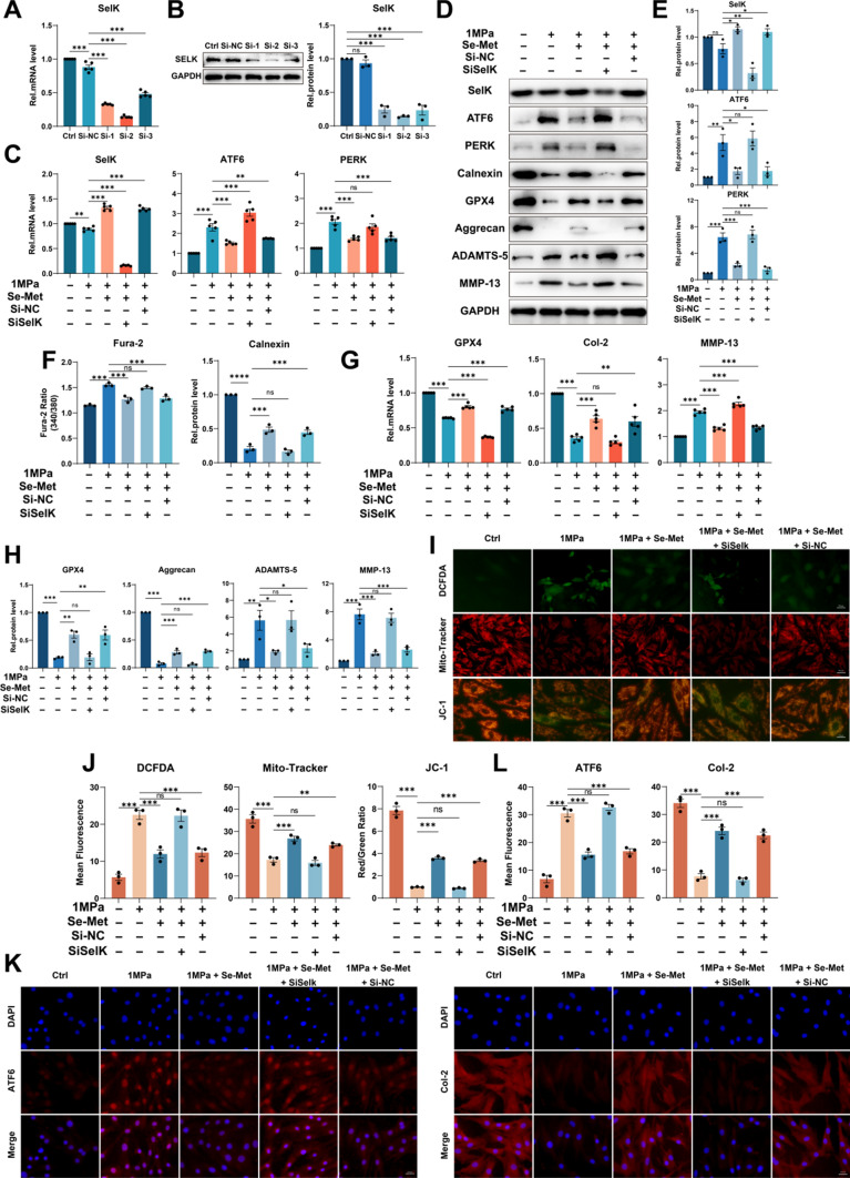
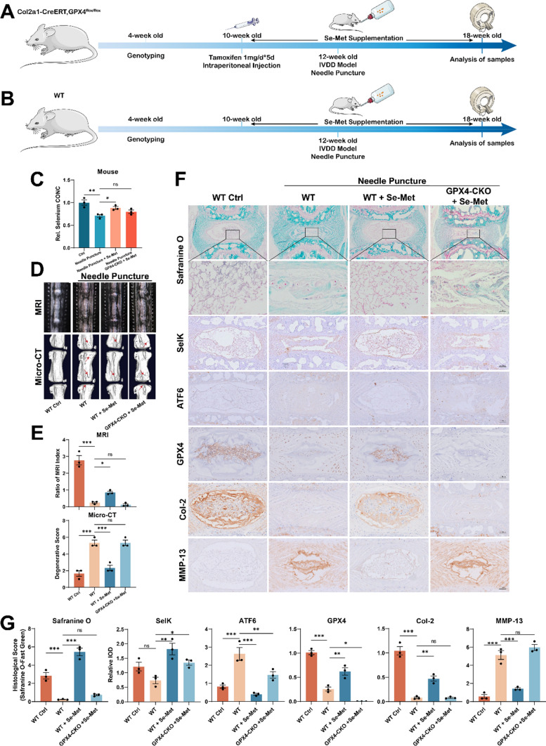
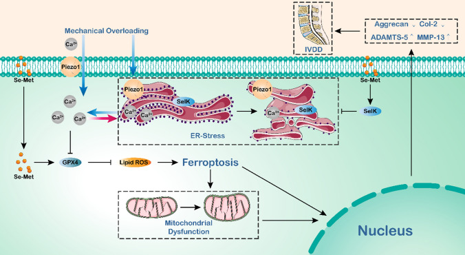
Intervertebral disc degeneration (IVDD) is one of the most prevalent spinal degenerative disorders and imposes places heavy medical and economic burdens on individuals and society. Mechanical overloading applied to the intervertebral disc (IVD) has been widely recognized as an important cause of IVDD. Mechanical overloading-induced chondrocyte ferroptosis was reported, but the potential association between ferroptosis and mechanical overloading remains to be illustrated in nucleus pulposus (NP) cells. In this study, we discovered that excessive mechanical loading induced ferroptosis and endoplasmic reticulum (ER) stress, which were detected by mitochondria and associated markers, by increasing the intracellular free Ca2+ level through the Piezo1 ion channel localized on the plasma membrane and ER membrane in NP cells. Besides, we proposed that intracellular free Ca2+ level elevation and the activation of ER stress are positive feedback processes that promote each other, consistent with the results that the level of ER stress in coccygeal discs of aged Piezo1-CKO mice were significantly lower than that of aged WT mice. Then, we confirmed that selenium supplementation decreased intracellular free Ca2+ level by mitigating ER stress through upregulating Selenoprotein K (SelK) expression. Besides, ferroptosis caused by the impaired production and function of Glutathione peroxidase 4 (GPX4) due to mechanical overloading-induced calcium overload could be improved by selenium supplementation through Se-GPX4 axis and Se-SelK axis in vivo and in vitro, eventually presenting the stabilization of the extracellular matrix (ECM). Our findings reveal the important role of ferroptosis in mechanical overloading-induced IVDD, and selenium supplementation promotes significance to attenuate ferroptosis and thus alleviates IVDD, which might provide insights into potential therapeutic interventions for IVDD.

Keywords: Endoplasmic reticulum stress; Ferroptosis; GPX4; Mechanical stress; SelK; Selenium.


Conflict of interest statement

The authors have no relevant financial or non-financial interests to disclose.

Figures

Fig. 1
Fig. 1
Excessive mechanical loading induces ferroptosis and ER stress in NP cells. A Typical human MR T2-weighted images of grade II (left panel) and grade IV (right panel) IVD. B General view of NP from donors who accepted NP discectomy surgery. The Safranine O-Fast Green staining of NP from human degenerated disc. The IHC staining of GPX4 and ACSL4 in human NP sections. Scale bar: 100 μm. C Microarray heatmap of rat NP cells from the control group and 1 MPa group (n = 3 for each group). D GO enrichment analysis of the indicated groups. E The IHC staining of ATF6 in human NP sections and the quantitative analysis. Scale bar: 100 μm. F The Western blotting analysis for GPX4 and ATF6 of NP tissues from humans and mice. G Representative TEM images and quantitative analysis of rat NP cells from the control group and 1 MPa group. Arrows show shrunken mitochondria and swollen ER. Scale bars: 5 μm (Low field), 1 μm (High field). H The Western blotting for ACSL4, GPX4, and Bip and quantification analysis (n = 3 for each group). I The qPCR of ACSL4, GPX4 (n = 4 for each group). J The Safranine O-Fast Green staining of coccygeal discs of WT mice and GPX4-CKO mice. Scale bar: 50 μm. Results were shown as mean ± SEM. *p < 0.05, ***p < 0.001
Fig. 2
Fig. 2
Excessive mechanical loading induces ferroptosis and ER stress by increasing the intracellular free Ca2+ level through the Piezo1 ion channel localized on the plasma membrane and ER membrane. A The qPCR of Piezo1 of the groups in this Figure (n = 3 for each group). B The Western blotting analysis for Piezo1 (n = 3 for each group). C The Fura-2 AM to detect intracellular free Ca2+ level (n = 3 for each group). D The qPCR of Calnexin, Bip, ACSL4, GPX4 and Col-2 (n = 3 for each group). E The Western blotting analysis for Calnexin, Bip, ACSL4, GPX4 and Col-2. F Quantification of Western blotting analysis (n = 3 for each group). GI DCFDA assays for ROS level and Mito-Tracker and JC-1 for mitochondrial membrane potential. Scale bar: 50 or 25 μm. J Quantitative analysis for DCFDA, Mito-Tracker and JC-1 (n = 3 for each group). K The IHC staining of Piezo1 and ATF6 for WT and Piezo1-CKO mice. Scale bar: 250 μm. L Quantitative analysis for the IHC staining of ATF6 (n = 3 for each group). Results were shown as mean ± SEM. ns not significant, *p < 0.05, **p < 0.01, ***p < 0.001
Fig. 3
Fig. 3
Suppression of ER stress attenuates mechanical overloading-induced ferroptosis by alleviating Ca2+ outflow from ER. A The qPCR of Bip, PERK, Calnexin, ACSL4 and GPX4 of the groups in this Figure (n = 3 for each group). B The Western blotting analysis for Bip, PERK, Calnexin, ACSL4, GPX4, Col-2 and MMP-13. C Quantification of Western blotting analysis (n = 3 for each group). D The Fura-2 AM to detect intracellular free Ca2+ level (n = 3 for each group). EG DCFDA assays for ROS level and Mito-Tracker and JC-1 for mitochondrial membrane potential. Scale bar: 50 or 25 μm. HI Quantitative analysis for DCFDA, Mito-Tracker, and JC-1 (n = 3 for each group). J The qPCR of Col-2 and MMP-13 of the groups in this Figure (n = 3 for each group). K Quantification of Western blotting analysis(n = 3 for each group). Results were shown as mean ± SEM. *p < 0.05, **p < 0.01, ***p < 0.001
Fig. 4
Fig. 4
Selenium supplementation protects NP cells from ferroptosis by enhancing GPX4 expression. A The concentration of selenium in NP tissues from humans (n = 3 for each group). B The concentration of selenium of NP cells (n = 3 for each group). C Microarray heatmap of rat NP cells from the 1 MPa group, and 1 MPa + Se-Met group (n = 3 for each group). D GO enrichment analysis of the indicated groups. E Representative TEM images and quantitative analysis of rat NP cells from 1 MPa group and 1 MPa + Se-Met group. Arrows show shrunken mitochondria and swollen ER. Scale bars: 2 μm (Low field), 1 μm (High field). F The qPCR of GPX4 of the groups in this Figure (n = 5 for each group). G The Western blotting analysis for GPX4 (n = 3 for each group). H DCFDA assays for ROS level and Mito-Tracker and JC-1 for mitochondrial membrane potential. Scale bar: 50 or 25 μm. I Quantitative analysis of mean fluorescence (n = 3 for each group). J The immunofluorescence of Col-2. Scale bar: 25 μm. K The qPCR of Aggrecan, Col-2, ADAMTS-5, and MMP-13 of the groups in this Figure (n = 5 for each group). L The Western blotting analysis for Aggrecan, Col-2, ADAMTS-5, and MMP-13. M Quantification of Western blotting analysis (n = 3 for each group). Results were shown as mean ± SEM. ns not significant, *p < 0.05, **p < 0.01, ***p < 0.001
Fig. 5
Fig. 5
Selenium supplementation mitigates ER stress through upregulating SELK expression. A The qPCR of SelK of the groups in this Figure (n = 5 for each group). B The Western blotting analysis for SelK (n = 3 for each group). C The qPCR of SelK, ATF6 and PERK of the groups in this Figure (n = 5 for each group). D The Western blotting analysis for SelK, ATF6, PERK, Calnexin, GPX4, Aggrecan, ADAMTS-5 and MMP-13. E Quantification of Western blotting analysis for SelK, ATF6 and PERK (n = 3 for each group). F The Fura-2 AM to detect intracellular free Ca2+ level (n = 3 for each group). G The qPCR of GPX4, Col-2 and MMP-13 of the groups in this Figure (n = 5 for each group). H Quantification of Western blotting analysis for GPX4, Aggrecan, ADAMTS-5 and MMP-13 (n = 3 for each group). I DCFDA assays for ROS level and Mito-Tracker and JC-1 for mitochondrial membrane potential. Scale bar: 50 or 25 μm. J Quantitative analysis of mean fluorescence (n = 3 for each group). K The immunofluorescence of ATF6 and Col-2. Scale bar: 25 μm. L Quantitative analysis of mean fluorescence (n = 3 for each group). Results were shown as mean ± SEM. ns not significant, *p < 0.05, **p < 0.01, ***p < 0.001
Fig. 6
Fig. 6
Selenium supplementation alleviates the progression of IVDD in mice. A Flowchart of animal experiment on GPX4-CKO mice. B Flowchart of animal experiment on WT mice. C The concentration of selenium of NP tissues from mice (n = 3 for each group). D The MRI and Micro-CT for coccygeal discs of mice. E Quantitative analysis of MRI and Micro-CT (n = 3 for each group). F The Safranine O-Fast Green staining and Immunohistochemical assay of SelK, ATF6, GPX4, Col2 and MMP-13. Scale bars: 50 or 250 μm. G Quantitative analysis of the Safranine O-Fast Green staining and Immunohistochemical assay (n = 3 for each group). Results were shown as mean ± SEM. ns not significant, *p < 0.05, **p < 0.01, ***p < 0.001
Fig. 7
Fig. 7
Schematic diagram of Selenium-SelK-GPX4 axis protects nucleus pulposus cells from mechanical overloading-induced ferroptosis

References

    1. Nava MM, et al. Heterochromatin-driven nuclear softening protects the genome against mechanical stress-induced damage. Cell. 2020;181:800–817.e822. doi: 10.1016/j.cell.2020.03.052. - DOI - PMC - PubMed
    1. Scott A, et al. High strain mechanical loading rapidly induces tendon apoptosis: an ex vivo rat tibialis anterior model. Br J Sports Med. 2005;39:e25. doi: 10.1136/bjsm.2004.015164. - DOI - PMC - PubMed
    1. Zhang X, et al. Mechanical stress regulates autophagic flux to affect apoptosis after spinal cord injury. J Cell Mol Med. 2020;24:12765–12776. doi: 10.1111/jcmm.15863. - DOI - PMC - PubMed
    1. Knezevic NN, Candido KD, Vlaeyen JWS, Van Zundert J, Cohen SP. Low back pain. Lancet. 2021;398:78–92. doi: 10.1016/S0140-6736(21)00733-9. - DOI - PubMed
    1. Ding SL, et al. Excessive mechanical strain accelerates intervertebral disc degeneration by disrupting intrinsic circadian rhythm. Exp Mol Med. 2021;53:1911–1923. doi: 10.1038/s12276-021-00716-6. - DOI - PMC - PubMed

LinkOut - more resources