Review
. 2024 Feb;55(2):311-323.
doi: 10.1161/STROKEAHA.123.043630. Epub 2024 Jan 22.

Epidemiology, Pathophysiology, and Imaging of Atherosclerotic Intracranial Disease

Affiliations
Review

Epidemiology, Pathophysiology, and Imaging of Atherosclerotic Intracranial Disease

Li Hui Chen et al. Stroke. 2024 Feb.

Abstract

Intracranial atherosclerotic disease (ICAD) is one of the most common causes of stroke worldwide. Among people with stroke, those of East Asia descent and non-White populations in the United States have a higher burden of ICAD-related stroke compared with Whites of European descent. Disparities in the prevalence of asymptomatic ICAD are less marked than with symptomatic ICAD. In addition to stroke, ICAD increases the risk of dementia and cognitive decline, magnifying ICAD societal burden. The risk of stroke recurrence among patients with ICAD-related stroke is the highest among those with confirmed stroke and stenosis ≥70%. In fact, the 1-year recurrent stroke rate of >20% among those with stenosis >70% is one of the highest rates among common causes of stroke. The mechanisms by which ICAD causes stroke include plaque rupture with in situ thrombosis and occlusion or artery-to-artery embolization, hemodynamic injury, and branch occlusive disease. The risk of stroke recurrence varies by the presumed underlying mechanism of stroke, but whether techniques such as quantitative magnetic resonance angiography, computed tomographic angiography, magnetic resonance perfusion, or transcranial Doppler can help with risk stratification beyond the degree of stenosis is less clear. The diagnosis of ICAD is heavily reliant on lumen-based studies, such as computed tomographic angiography, magnetic resonance angiography, or digital subtraction angiography, but newer technologies, such as high-resolution vessel wall magnetic resonance imaging, can help distinguish ICAD from stenosing arteriopathies.

Keywords: arteries; cerebral infarction; demography; intracranial arteriosclerosis; stroke.


Conflict of interest statement

Disclosures Dr Gutierrez received compensation for contributions to an up-to-date article on intracranial atherosclerosis and as an expert witness. The other authors report no conflicts.

Figures

Figure 1
Figure 1
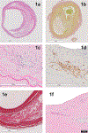
Intracranial atherosclerosis Intracranial atherosclerosis consist of cholesterol deposition in the intima coalescing into an atheroma (Figure 1a, H&E stain and Figure 1b, EVG stain). The atheroma is surrounded by intense intima proliferation and fibrosis, usually in an eccentric causing various degrees of luminal stenosis (Figure 1a and b, lumen denoted by the letter L). Foam cells consist of macrophages that swallow fatty particles and appear round with an eccentric nuclei (Figure 1c, blue arrow). Foam cells stain positive for CD68 (Figure 1d, +CD68 cells appeared brown). The fibrous cap (Figure 1e, doubled arrow head marking the thickness of the fibrous cap) separates the atheroma from the lumen (L). A thinner fibrous cap is thought to increase the vulnerability of the plaque to rupture. Other high-risk features off plaque include intracranial hemorrhage and neovascularization (Figure 1f show evidence of a small vessel inside the plaque). Neovascularization should not be confused with vasa vasorum, which surround the adventitia in the most proximal parts off the circle of Willis but rarely beyond the carotid or vertebral arteries.
Figure 2
Figure 2
Pathophysiology of intracranial atherosclerotic disease Intracranial atherosclerotic stenosis can cause stroke mainly by three mechanisms, which can be overlapping. 1) Plaque rupture: it occurs when the endothelium is breached, usually with a thin fibrous cap, and thrombogenic material is exposed to the blood stream. The resulting thrombus formation on the plaque luminal surface can grow and occlude the artery at the level off the stenosis, or it can detach entirely or partially causing distal embolization. 2) Hemodynamic effect: Plaques can continually grow and increase the degree of stenosis. Progressive stenosis can reduce forward flow, causing hypoperfusion, which can be exacerbated in the setting of hypovolemia or hypotension. 3) Branch occlusive disease: Atheromas are accompanied by various degree off intima proliferation and increased ground substance, which might narrow or occlude the ostia of penetrating arteries branching of the parent artery and manifest radiographically and clinically as a lacunar stroke.
Figure 3
Figure 3
Lumen-based study of intracranial atherosclerotic stenosis. Intracranial atherosclerotic stenosis is diagnosed mostly by lumen-based studies. Patients with high-degree CAD can have stroke via hemodynamic failure (Figure 3a and c showing a brain CT with a flow gap in the proximal left m1 and CT perfusion showing hypoperfusion). Similarly, ICAD can impinge in the origin of penetrating arteries and cause a lacunar infarct (Figure 3c shows high degree stenosis of the right M1 with a co-existing acute infarct shown in DWI images in figure 3d). Lumen-based studies such as CTA, time-of-flight MRA (Figure 3e showing flow artifact exaggerating the degree of stenosis) or DSA (Figure 3f shows the same stenosis as in Figure 3e but delimits better the vessel contours avoiding MRA time-of-flight flow artifact) are the most common diagnostic methods of ICAD.
Figure 4
Figure 4
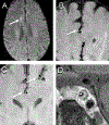
High resolution vessel wall MRI examples Man in his 60’s with scatted foci of restricted diffusion involving the right frontal lobe centrum semiovale (Figure 4A, arrow, DWI sequence). There is moderate focal narrowing of the right pericallosal artery (Figure 4B, right ACA A3 segment pointed by the arrow) due to eccentric wall thickening compatible with atherosclerotic plaque (Figure 3C, arrows). High-resolution vessel wall MRI can help identify atherosclerotic plaque components (long white arrow, contrast-enhancing fibrous cap; short white arrow, hypointense lipid core) shown at a cavernous segment of a left ICA.

References

    1. Gorelick PB, Wong KS, Bae H-J and Pandey DKJS. Large artery intracranial occlusive disease: a large worldwide burden but a relatively neglected frontier. 2008;39:2396–2399. - PubMed
    1. White H, Boden-Albala B, Wang C, Elkind MS, Rundek T, Wright CB and Sacco RLJC. Ischemic stroke subtype incidence among whites, blacks, and Hispanics: the Northern Manhattan Study. 2005;111:1327–1331. - PubMed
    1. Sharma VK, Tsivgoulis G, Teoh HL, Ong BK, Chan BPJJoS and Diseases C. Stroke risk factors and outcomes among various Asian ethnic groups in Singapore. 2012;21:299–304. - PubMed
    1. Tsivgoulis G, Vadikolias K, Heliopoulos I, Katsibari C, Voumvourakis K, Tsakaldimi S, Boutati E, Vasdekis SN, Athanasiadis D and Al-Attas OSJJoN. Prevalence of symptomatic intracranial atherosclerosis in Caucasians: a prospective, multicenter, transcranial Doppler study. 2014;24:11–17. - PubMed
    1. Mazighi M, Labreuche J, Gongora-Rivera F, Duyckaerts C, Hauw J-J and Amarenco PJS. Autopsy prevalence of intracranial atherosclerosis in patients with fatal stroke. 2008;39:1142–1147. - PubMed