. 2024 Jan 22;15(1):81.
doi: 10.1038/s41419-024-06440-z.

RBL2 represses the transcriptional activity of Multicilin to inhibit multiciliogenesis

Affiliations

RBL2 represses the transcriptional activity of Multicilin to inhibit multiciliogenesis

Erik J Quiroz et al. Cell Death Dis. .

Abstract

A core pathophysiologic feature underlying many respiratory diseases is multiciliated cell dysfunction, leading to inadequate mucociliary clearance. Due to the prevalence and highly variable etiology of mucociliary dysfunction in respiratory diseases, it is critical to understand the mechanisms controlling multiciliogenesis that may be targeted to restore functional mucociliary clearance. Multicilin, in a complex with E2F4, is necessary and sufficient to drive multiciliogenesis in airway epithelia, however this does not apply to all cell types, nor does it occur evenly across all cells in the same cell population. In this study we further investigated how co-factors regulate the ability of Multicilin to drive multiciliogenesis. Combining data in mouse embryonic fibroblasts and human bronchial epithelial cells, we identify RBL2 as a repressor of the transcriptional activity of Multicilin. Knockdown of RBL2 in submerged cultures or phosphorylation of RBL2 in response to apical air exposure, in the presence of Multicilin, allows multiciliogenesis to progress. These data demonstrate a dynamic interaction between RBL2 and Multicilin that regulates the capacity of cells to differentiate and multiciliate. Identification of this mechanism has important implications for facilitating MCC differentiation in diseases with impaired mucociliary clearance.


Conflict of interest statement

The authors declare no competing interests.

Figures

Fig. 1
Fig. 1. Multicilin physically interacts with Rbl2 in mouse embryonic fibroblasts.
ac Schematics of the adenoviral constructs for FLAG-MCI (a), FLAG-MCI+E2f4 (b), and FLAG-MCI+E2f4VP16 (c). d Normalized spectral abundance factor (NSAF) for the top 10 most abundant proteins co-immunoprecipitating with FLAG-MCI relative to FLAG-MCI+E2f4VP16 (see Supplementary Table S1). e Representative silver stain of total protein lysate, the red arrowhead indicates 3xFLAG-MCI. f Representative western blot analysis for Rbl1, Rbl2, and FLAG for total protein (top) and co-immunoprecipitations (Co-IP, bottom) with FLAG from non-infected mouse embryonic fibroblasts (MEFs) and MEFs infected with Ad5 FLAG-MCI, FLAG-MCI+E2f4 and FLAG-MCI+E2f4VP16.
Fig. 2
Fig. 2. Rbl2 depletion increases the transcriptional activity of Multicilin stimulating multiciliogenesis in fibroblasts.
a Representative western blot for RBL2, RBL1 and centriole biogenesis proteins STIL and DEUP1, comparing FLAG-Multicilin in the presence of a non-targeting siRNA (siCTL) or siRNA targeting Rbl1 (siRbl1), Rbl2 (siRbl2), or both siRbl1 and siRbl2 (siRbl1/2). b qRT-PCR for MCC genes Mcidas, Ccno and Deup1 comparing FLAG-Multicilin with either siRbl2 or E2f4VP16 relative to siCTL (Data expressed as mean ± SEM., N = 2 experimental replicates, normalized to Gapdh). c Confocal images comparing FLAG-Multicilin in the presence of either siCTL or siRbl2 stained for centriole biogenesis markers DEUP1 (red), SAS-6 (green) and nuclei are counterstained with DAPI (blue). Scale bars = 20 µm. d Representative western blot comparing FLAG-Multicilin in the presence siCTL, siRbl2, or E2f4VP16 for RBL2, RBL1, FLAG, axonemal proteins CCDC39 and RSPH9 and γ-Tubulin (loading control). e Confocal images for cilia marker ARL13B (green) and basal body marker CEP164 (red) nuclei are counterstained with DAPI (blue). Cilia are quantified per cell, data represents mean ± SD, N = 3, **** P < 0.0001, unpaired t-test. Scale bar = 10 µm.
Fig. 3
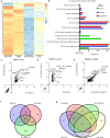
Fig. 3. Transcriptome profiling confirms induction of multiciliogenesis in the presence of siRbl2 or E2f4VP16 compared to Multicilin alone.
a Unsupervised heatmap of RNA-seq data for MCC-associated genes comparing non-infected MEFs and FLAG-MCI MEFs with either siCTL, siRbl2 or E2f4VP16. b Gene ontology (GO) analysis of differentially expressed genes (DEGs) (Supplementary Table S3) comparing non-infected MEFs and FLAG-MCI MEFs with either siCTL, siRbl2 or E2f4VP16. GO terms include biological process (GO:BP) and cellular components (GO:CC). ce Scatter plots comparing significant DEGs in FLAG-MCI MEFs (relative to uninfected, absolute log2 fold change>2, adj. p < 0.5) for siRbl2 vs. siCTL (c), E2f4VP16 vs siCTL (d), and siRbl2 vs E2f4VP16 (e). f, g Venn diagrams representing significantly increased DEGs in FLAG-MCI relative to uninfected controls (log2 fold change>2, adj. p < 0.5, Supplementary Table S5) comparing siRbl2, siCTL and E2f4VP16 (f) and compared to known MCC-specific genes [37] (g). N = 3 experimental replicates for all RNAseq data.
Fig. 4
Fig. 4. RBL2 knockdown does not impact the efficiency of MCC differentiation in HBEC.
qRT-PCR comparing HBECs treated with shRNA-RBL2 (shRNA#1 (filled circles), shRNA#2 (open squares)) or a non-targeting control (NTC) at Day 14 if ALI culture for RBL2 (a), early MCC markers SAS6, Deup1 and TP73 (b) and differentiated epithelial cell markers FOXJ1 (MCC), CC10 (club), and Muc5AC (goblet) (c). Data is corrected for RPLP0 and normalized to NTC shRNA, N = 4–5 biological replicates each with n = 1–2 experimental replicates paired for each shRNA. d Representative western blot for shRNA#1 and NTC#1 at Day 14 ALI for RBL2 and MCC proteins CCDC39, TP73 and FOXJ1. e Quantification of protein expression from N = 3 biological replicates for each shRNA. f Quantification of cilia coverage at D14 ALI expressed as a ratio of area of total acetylated α-tubulin (ATUB, cilia) over total growth area (area F-Actin), N = 5–6 biological replicates each n = 1-2 experimental replicates. g Analysis of cilia beat frequency (CBF) at D14 ALI, N = 4–5 biological replicates. h Representative IF images of ATUB (green), scale bars represent 1 mm and 100 µm. Data is expressed as mean ± SEM and compared using two-tailed paired t-tests (ac, e) ratio-paired t-test (f) paired t-test (g) and *P < 0.05, ** P < 0.01, *** P < 0.001, **** P < 0.0001.
Fig. 5
Fig. 5. Air-liquid interface attenuates the interaction between RBL2-Multicilin.
a Representative western blot for total protein and immunoprecipitation with FLAG in FLAG-MCI induced HBECs either submerged (Sub) or ALI culture. b Representative western blot for total RBL2, phosphorylated serine 672 RBL2 (RBL2 S672) and MCC proteins TP73 and FOXJ1 at D0, D4 (Sub) and ALI D4. Protein quantification for total-RBL2 (c) and RBL2 S672:total RBL2 (d), N = 6 biological replicates. e Representative western blot for Multicilin-induced HBECs at Sub D4 in the presence of 5 µM DAPT or vehicle. f Protein quantification for DAPT treatment from N = 3, from 1-2 biological replicates and 1-2 experimental replicates. Data represents mean ± SEM and is compared using a paired t-test (c, d) and 2-way ANOVA and post-hoc Tukey’s multiple comparisons test (f).
Fig. 6
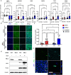
Fig. 6. RBL2 inhibits the activity of Multicilin in HBECs.
a RT-qPCR for HBECs comparing nontargeting controls (shNTC#2, red) to RBL2 (shRBL2#2, blue) in the presence (DOX) or absence (Veh, H2O) of Multicilin induction. Gene expression is relative to RPLP0 and normalized to the NTC shRNA. N = 4 biological replicates, n = 2 experimental replicates (squares, triangles). b Representative IF images for FLAG (Green), TP73 (Cyan) and DNA (DAPI, blue) in HBECs. Scale bars represent 200 µm. c Quantification of TP73 positive cells. N = 3–4 biological replicates per shRNA d Representative western blot for RBL2, TP73 and FLAG in HBECs comparing shNTC#2 to shRBL2#2 in the presence (DOX) or absence (Veh) of Multicilin induction e Representative confocal images of basal bodies (Pericentrin, cyan) and cilia (acetylated α-tubulin, ATUB, green) with DNA counterstain (DAPI, blue) in shRBL2 with Multicilin induction. Scale bars represent 25 µm. Data represents mean ± SEM and is compared using a 2-way ANOVA and post-hoc Tukey’s multiple comparisons test (ac) with significance at *P < 0.05, **P < 0.01, ***P < 0.001, ****P < 0.0001.
Fig. 7
Fig. 7. Regulation of Multiciliogenesis and the activity of Multicilin by RBL2.
Schematic depicting the new mechanism of regulating MCI activity during multiciliogenesis. Unphosphorylated RBL2 binds to E2F4 and inhibits Multicilin-induction of MCC gene expression. Differentiation at an air-liquid interface and inhibition of NOTCH signaling both phosphorylate RBL2 and dissociate it from the E2F4/Multicilin complex, allowing for induction of MCC gene expression.

Update of

References

    1. Brooks ER, Wallingford JB. Multiciliated cells. Curr Biol. 2014;24:R973–82. doi: 10.1016/j.cub.2014.08.047. - DOI - PMC - PubMed
    1. Hoque M, Kim EN, Chen D, Li FQ, Takemaru KI. Essential Roles of Efferent Duct Multicilia in Male Fertility. Cells. 2022;11:341. doi: 10.3390/cells11030341. - DOI - PMC - PubMed
    1. Meunier A, Azimzadeh J. Multiciliated Cells in Animals. Cold Spring Harb Perspect Biol. 2016;8:a028233. doi: 10.1101/cshperspect.a028233. - DOI - PMC - PubMed
    1. Li Q, Han Z, Singh N, Terre B, Fame RM, Arif U, et al. Disruption of GMNC-MCIDAS multiciliogenesis program is critical in choroid plexus carcinoma development. Cell Death Differ. 2022;29:1596–610. doi: 10.1038/s41418-022-00950-z. - DOI - PMC - PubMed
    1. Roberson EC, Tran NK, Konjikusic MJ, Fitch RD, Gray RS, Wallingford JB. A comparative study of the turnover of multiciliated cells in the mouse trachea, oviduct, and brain. Dev Dyn. 2020;249:898–905. doi: 10.1002/dvdy.165. - DOI - PMC - PubMed

Publication types

Substances